
|
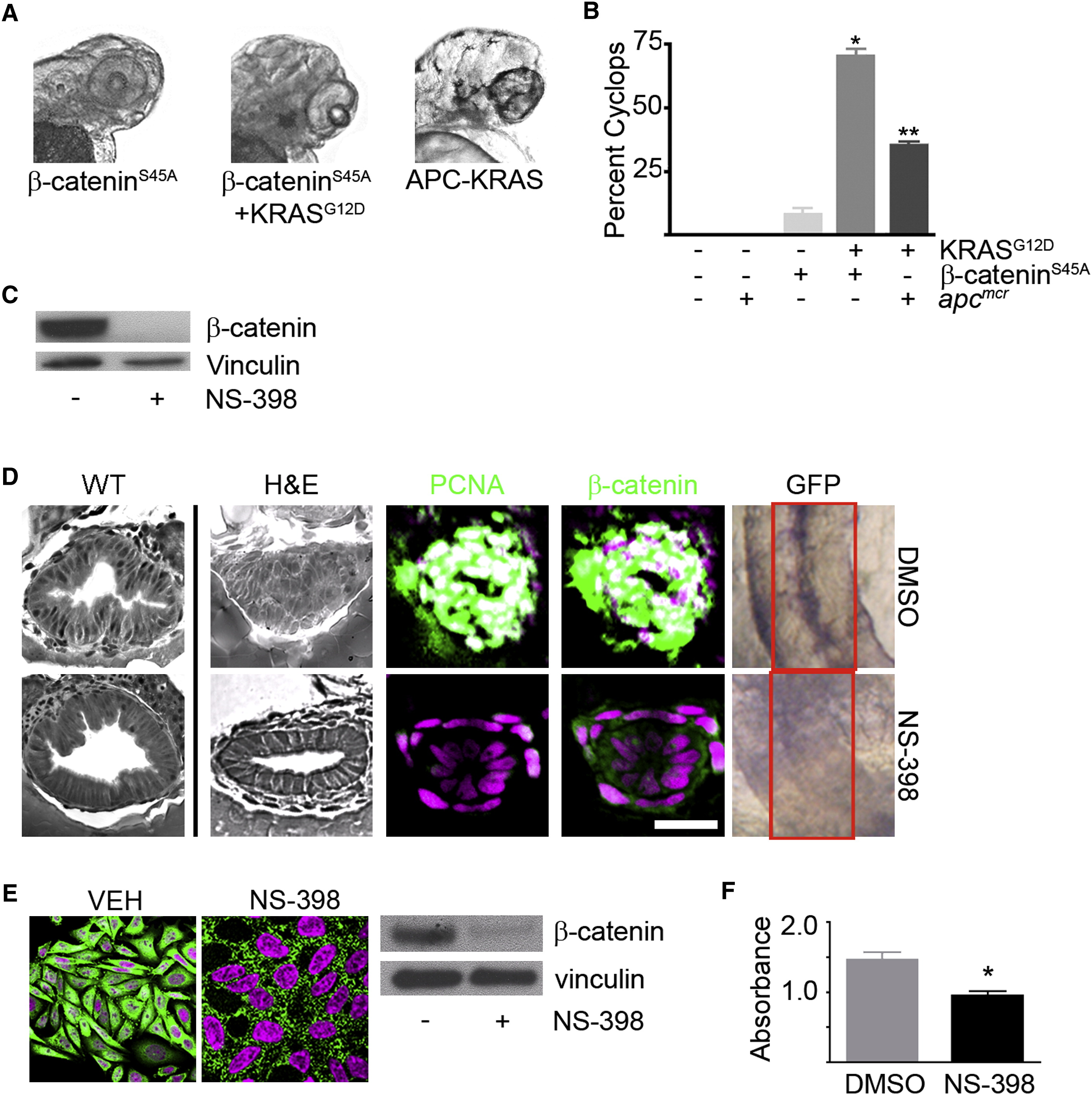
Fig. 5 KRAS-Mediated Intestinal Cell Proliferation following Loss of APC Requires β-Catenin
(A) Zebrafish embryos were injected with β-cateninS45A mRNA (left) along with KRAS mRNA (middle). Also shown is a representative image of the APC-KRAS embryo (right). At 72 hpf, the embryos were fixed and photographed.
(B) The percent cyclops was analyzed (*p < 0.01 WT versus β-catenin-KRAS, **p < 0.01 WT versus APC-KRAS).
(C) Protein was harvested from apcmcr embryos treated with DMSO or NS-398 and subjected to western blot analysis for β-catenin (top) or vinculin (bottom).
(D) Wild-type uninjected or apcmcr embryos injected with KRAS mRNA treated with VEH (top) or NS-398 (bottom) were stained by H&E (WT, left; APC-KRAS, right) and for DNA (magenta), PCNA (green), and β-catenin (green). TOPGFP-APCmo-KRAS embryos were stained for GFP expression (purple). Boxes indicate the intestine.
(E) SW-480 cells treated with DMSO or NS-398 were stained for DNA (magenta) and β-catenin (green). Protein lysates were subjected to western blot analysis for β-catenin (top) and vinculin (bottom).
(F) Cells were subjected to MTT analysis (*p < 0.05 versus DMSO; error bars, SEM).
Overlapping expression is shown in white. All images were captured using the same exposure and represent at least three independent experiments. Scale bar, 10 μm.
Reprinted from Cell, 137(4), Phelps, R.A., Chidester, S., Dehghanizadeh, S., Phelps, J., Sandoval, I.T., Rai, K., Broadbent, T., Sarkar, S., Burt, R.W., and Jones, D.A., A two-step model for colon adenoma initiation and progression caused by APC loss, 623-634, Copyright (2009) with permission from Elsevier. Full text @ Cell