
|
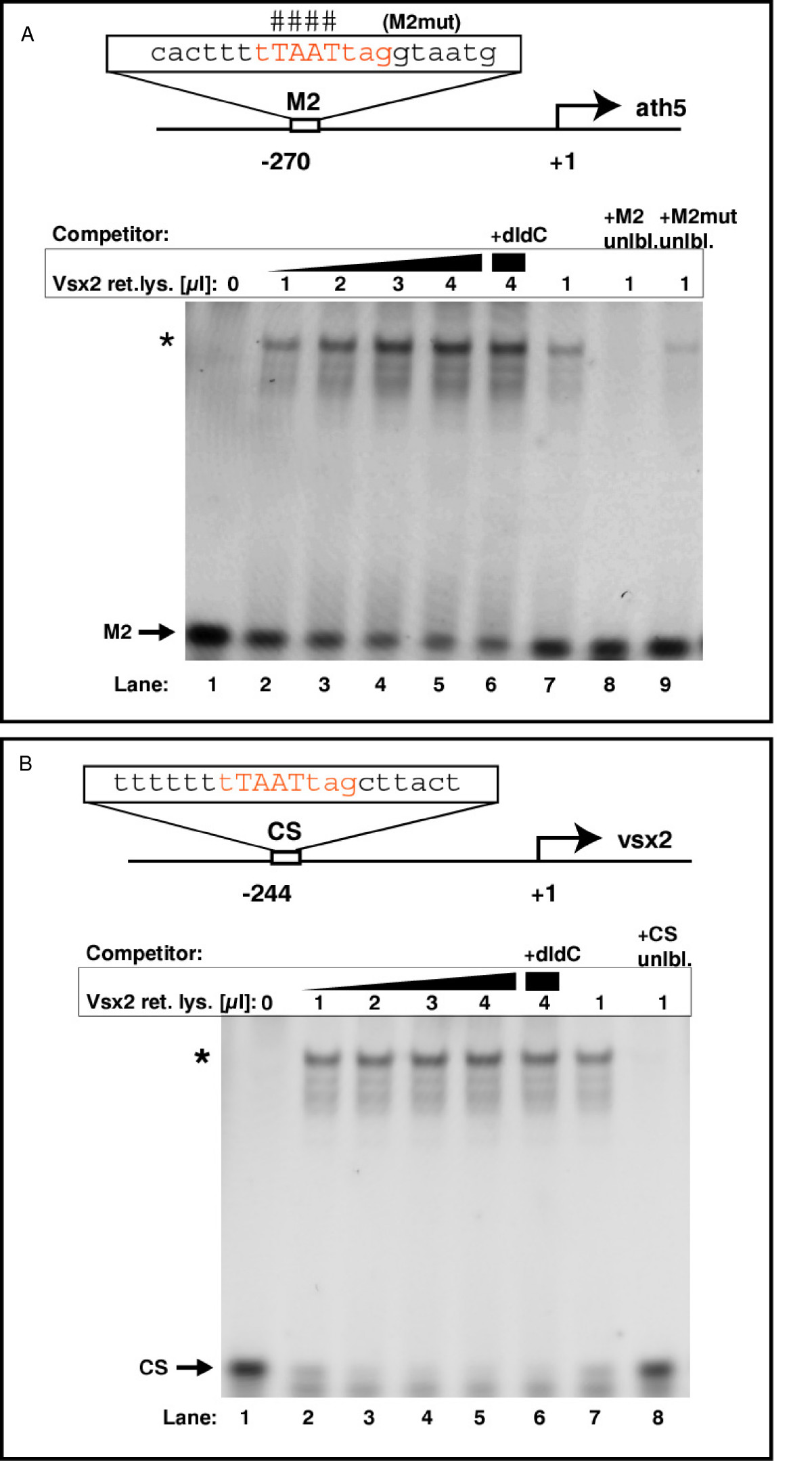
Fig. 10 Vsx2 binds to ath5 and vsx2 promoter sequences in vitro. (A)In vitro translated Vsx2 protein was incubated with a Cy5-end-labeled probe against the ath5 promoter fragment (M2) with or without competitors. The binding sequence core of M2 is shown in red. Lanes 2–5: addition of increasing amounts of Vsx2 protein shifts cumulative amounts of M2 oligo (asterisk; band shift). In vitro translated luciferase protein served as negative control and induced no band shift (lane 1). Addition of the unspecific competitor poly(dI-dC) (lane 6) did not reduce binding. Lanes 7–9: band shift of labelled M2 oligo by Vsx2 was challenged by adding specific competitors. Addition of excess unlabeled M2 oligo led to out-competition of the labelled M2 oligo (lane 8 versus lane 7). Addition of excess unlabeled mutated M2 oligo could compete only weakly with binding (lane 9). The bases mutated in this oligo are indicated by the hash symbols above the binding sequence core. (B) In vitro translated Vsx2 protein was incubated with a Cy5-end-labeled probe against the vsx2 promoter fragment (CS) with or without competitors. The binding sequence core of CS is shown in red. Lanes 2–5: addition of increasing amounts of Vsx2 protein shifts cumulative amounts of CS oligo (asterisk; band shift). In vitro translated luciferase protein served as negative control and induced no band shift (lane 1). Addition of the unspecific competitor poly(dI-dC) (lane 6) did not reduce binding significantly. Lanes 7–8: addition of excess unlabeled CS oligo led to out-competition of the labelled CS oligo.