
|
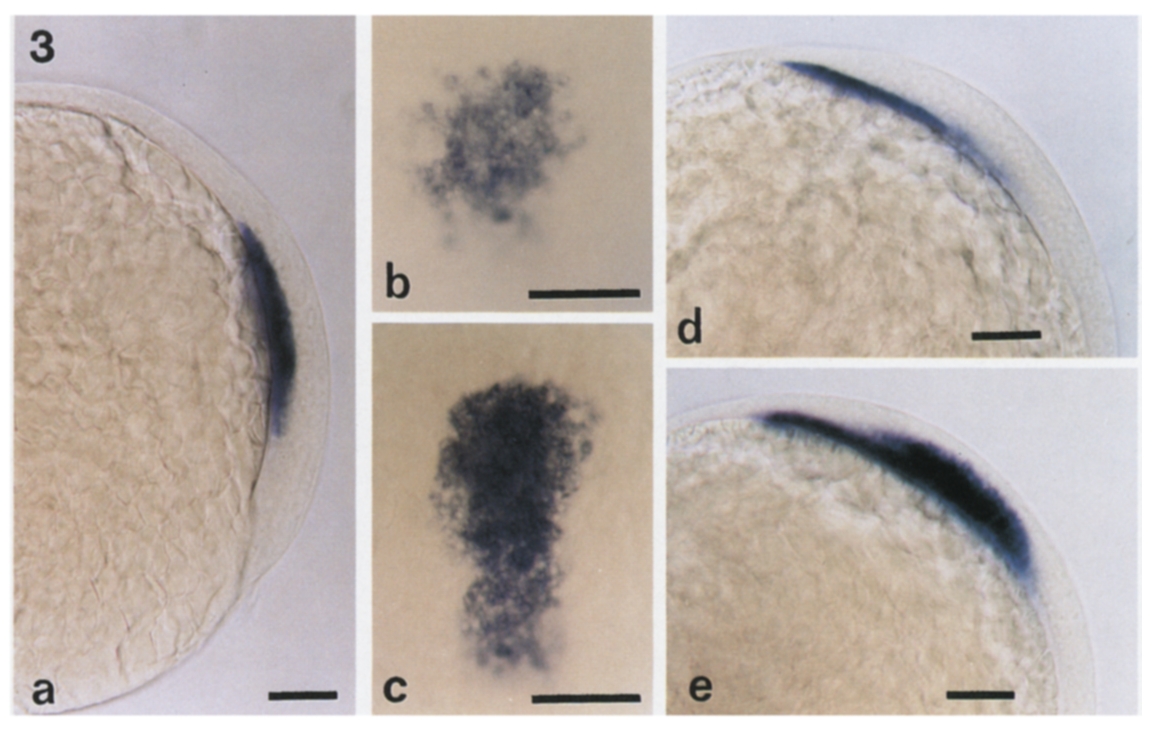
Fig. 3 Localization of gsc RNA in cyclops (cycb16) mutant embryos. (a) Side view of a whole mount cyclops embryo at 70% epiboly. Expression of gsc in the mesendoderm is reduced compared to wild-type control (Fig. 2a) (animal pole up; dorsal, right). (b) Dorsal view of the same embryo. gsc is confined to the most anterior part of the mesendodermal layer and encompasses only a portion of the wild-type gsc territory shown in (c) (anterior, top). (c) Dorsal view of a wild-type sibling embryo at 70% epiboly, showing gsc expression in the entire axial mesendoderm (compare to (b); see also Fig. 2a) (anterior, top). (d) Side view of a cyclops embryo at 95% epiboly, gsc expression is not detected in the ectoderm and is reduced in the mesodermal territory compared to wild-type expression shown in (e) (anterior, upper left). (e) Wild-type sibling embryo at 95% epiboly. Scale bars, 100 μm. Phenotypically mutant embryos were approximately one-quarter (23.4% = 55/235) of the total, as expected for simple Mendelian segregation.
Reprinted from Developmental Biology, 164, Thisse, C., Thisse, B., Halpern, M.E., and Postlethwait, J.H., Goosecoid expression in neurectoderm and mesendoderm is disrupted in zebrafish cyclops gastrulas, 420-429, Copyright (1994) with permission from Elsevier. Full text @ Dev. Biol.