
|
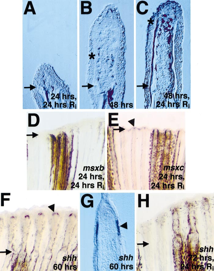
Fig. 6 Fgfr1 inhibition blocks outgrowth and reduces msxb/c and shh expression in ongoing fin regenerates. (A) Section of amputated fin from fish allowed to regenerate for 24 h before a 24-h treatment with Ri. (B) Section of untreated fin at 48 h postamputation. (C) Section of fin from fish allowed to regenerate for 48 h before treatment with Ri. Note that little outgrowth occurred during Fgfr1 inhibition. By comparing (B) and (C), it is apparent that, during Ri treatment, melanocyte migration and bone matrix deposition (still inconspicuous in (B)) continued despite the lack of outgrowth. Asterisks denote distalmost points of bone deposition (C) and pigment cell localization (B, C), and arrows demarcate the amputation plane in each photograph. (D, E) Whole-mount views of fin allowed to regenerate for 24 h, treated with Ri, and assessed for msxb (D) or msxc (E) expression. Note that Ri treatment removed (D) or diminished (E; arrowhead marks signal) established expression (compare with Figs. 2E and 2I). (F) shh was expressed at 60 h postamputation in dual fin ray domains. (G) Section through fin shown in (F) indicates bilateral shh expression domains in the basal layer of the epidermis. (H) Fin allowed to regenerate for 72 h, treated with Ri, and stained for shh mRNA expression. shh transcripts were undetectable following Ri application. Original magnification in (A–C, G) was 250x and in (D, E, H) 50x.
Reprinted from Developmental Biology, 222(2), Poss, K.D., Shen, J., Nechiporuk, A., McMahon, G., Thisse, B., Thisse, C., and Keating, M.T., Roles for Fgf signaling during zebrafish fin regeneration, 347-358, Copyright (2000) with permission from Elsevier. Full text @ Dev. Biol.