
|
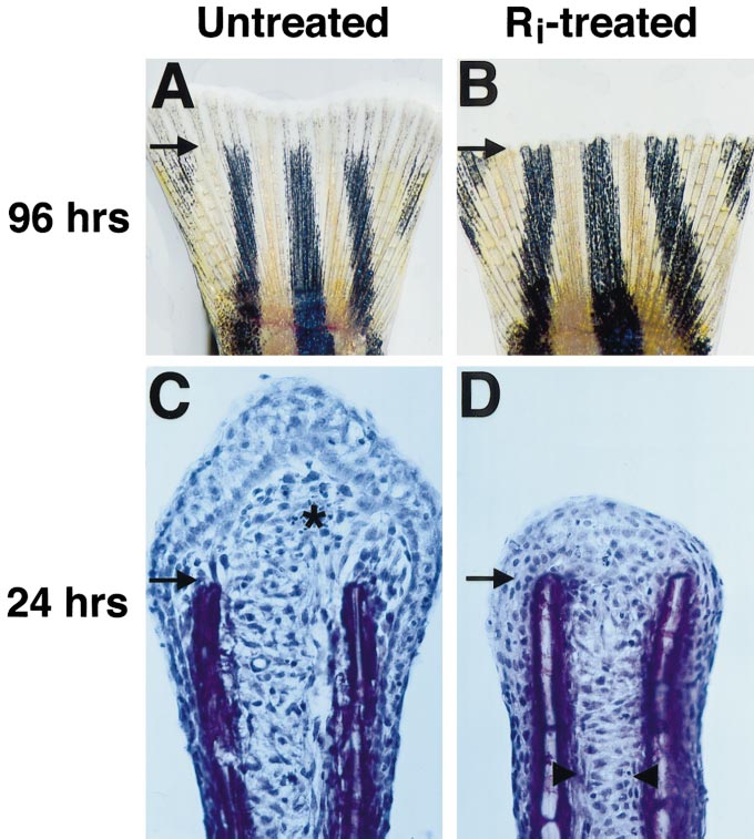
Fig. 3 Fgfr1 inhibition blocks fin regeneration. (A) Fin from untreated fish at 96 h after amputation, showing normal regrowth and new segmentation. Arrows demarcate amputation plane in each photograph. (B) Fin from fish treated with Ri for 96 h immediately following amputation. These fins showed no new growth. Here, the amputated edge appears saw-toothed due to the retraction of tissue between rays. (C) Hematoxylin stain of 24-h fin regenerate section from untreated fish (asterisk denotes new blastema). (D) Fin regenerate section from fish treated with Ri for 24 h. Note the lack of blastema. However, Ri-treated fin regenerates showed mesenchymal disorganization (arrowheads mark boundary between organized and disorganized tissue), as well as longitudinal arrangement suggestive of migration. Original magnification in (A, B) was 20x and in (C, D) 400x.
Reprinted from Developmental Biology, 222(2), Poss, K.D., Shen, J., Nechiporuk, A., McMahon, G., Thisse, B., Thisse, C., and Keating, M.T., Roles for Fgf signaling during zebrafish fin regeneration, 347-358, Copyright (2000) with permission from Elsevier. Full text @ Dev. Biol.