
|
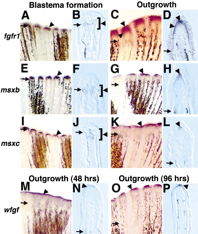
Fig. 2 Fgfr1, msxb, msxc, and wfgf are expressed in the fin regenerate during blastema formation (18–24 h postamputation) and/or regenerative outgrowth (48 h postamputation unless otherwise indicated). (A) Fgfr1 expression at 24 h postamputation (violet stains indicate positive signal, arrowhead). Arrows demarcate amputation plane in each photograph. (B) Longitudinal section (bottom, anterior; top, posterior) through an 18-h fin stained for fgfr1. Mesenchymal cells located between and just distal to hemirays faintly expressed fgfr1 (brackets outline region of expression). (C) Fgfr1 expression at 48 h postamputation. (D) Section through a 48-h fin regenerate stained for fgfr1 mRNA expression. Two different domains are evident: one in mesenchymal cells representing the distal blastema and one expressing bilaterally in the basal layer of the regeneration epidermis. (E) Msxb expression at 24 h postamputation. (F) Longitudinal section of 18-h fin regenerate assessed for msxb expression, indicating msxb-positive cells between and extending beyond hemirays. (G) msxb expression at 48 h postamputation. (H) Section of 48-h fin regenerate labeled for msxb demonstrates expression in the distal blastema. (I) msxc expression at 24 h postamputation. (J) Longitudinal section of 18-h fin indicates msxc-positive cells between hemirays. (K, L) Expression of msxc at 48 h was virtually identical to that of msxb in whole mounts (K) and sections (L). (M) wfgf expression at 48 h postamputation. (N) Longitudinal section of fin stained for wfgf expression at 48 h. Only the distalmost cells of the regeneration epidermis expressed wfgf. (O) wfgf expression at 96 h postamputation. (P) Section of 96-h regenerate assessed for wfgf expression, indicating an epidermal hybridization signal. Original magnification in whole mounts (A, C, E, G, I, K, M, O) was 50x and was 250x in sections (B, D, F, H, J, L, N, P).
Reprinted from Developmental Biology, 222(2), Poss, K.D., Shen, J., Nechiporuk, A., McMahon, G., Thisse, B., Thisse, C., and Keating, M.T., Roles for Fgf signaling during zebrafish fin regeneration, 347-358, Copyright (2000) with permission from Elsevier. Full text @ Dev. Biol.