
|
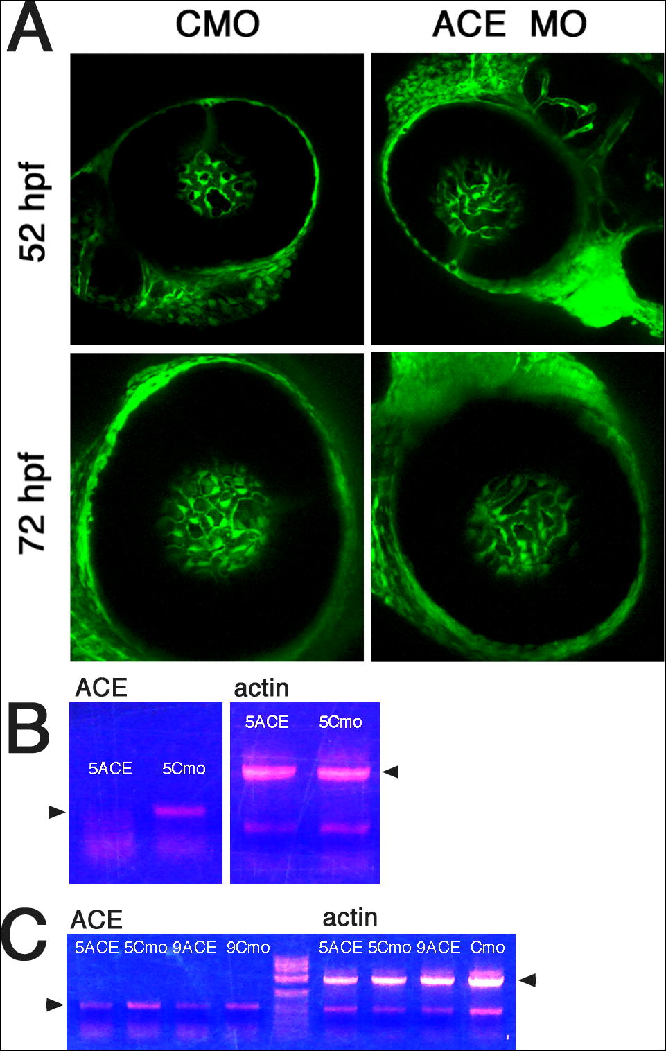
Fig. S1 Analysis of retinal vasculature in ACE knockdown animals. (A) Confocal images of retinae from flk-GFP transgenic animals treated with anti-ACE or control (CMO) morpholinos. Lateral views are provided of representative retinae from four independent experiments. (B) RT-PCR analysis of knockdown efficiency at 24 hpf. 5 μg/μl of morpholino was used in this experiment. (C) RT-PCR analysis of knockdown efficiency at 48 hpf. Two morpholino concentrations were used: 5 and 9 μg/μl. The amplification of actin mRNA was used to control the amount and the quality of RNA extracted from morphant and control animals. Arrowheads indicate relevant amplification products.
Reprinted from Mechanisms of Development, 126(5-6), Kitambi, S.S., McCulloch, K.J., Peterson, R.T., and Malicki, J.J., Small molecule screen for compounds that affect vascular development in the zebrafish retina, 464-477, Copyright (2009) with permission from Elsevier. Full text @ Mech. Dev.