
|
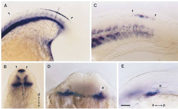
Fig. 6 snail2 expression pattern during late somitogenesis in the tail and in the otic vesicle region. (A) Expression pattern of snail2 in the posterior trunk and tail bud of an embryo at the 16-somite stage. On the dorsal side of the embryo (toward the top of the figure) snail2 staining in the neural crest extends 12 somites anteriorly from the tail bud (arrowheads). Ventrally, the staining is very strong in mesoderm. (B) An optical section through the tail region of the same embryo viewed at the level of the dashes in A (dorsal, top). snail2 transcripts appear in neural crests dorsally in the neural rod (between arrowheads) and in the medial and ventral part of the somite. Less intense staining occurs in muscle pioneer precursors located lateral to the notochord (n). (C) Tail of a 24-hr-old embryo. snail2 expression appears at the position of neural crest in 3 to 4 of the most recently formed somites (between arrowheads). (D) Optical section of a 24-hr-old embryo at the level of the otic vesicle (e) and (E) lateral view of the same embryo. snail2 transcripts accumulate just ventral and anterior to the otic vesicle. dorsoventral (d–v) and anteroposterior (a–p) axis. Scale bars, 25 μm (A), 40 μm (B–E).
Reprinted from Developmental Biology, 172, Thisse, C., Thisse, B., and Postlethwait, J.H., Expression of snail2, a second member of the zebrafish Snail family, in cephalic mesendoderm and presumptive neural crest of wild-type and spadetail mutant embryos, 86-99, Copyright (1995) with permission from Elsevier. Full text @ Dev. Biol.