
|
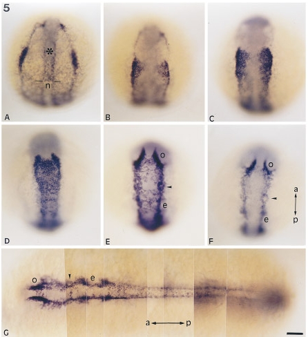
Fig. 5 snail2 expression during somitogenesis. (A–F) dorsal view of embryos at (A) 3-somite stage, (B) 5-somite stage, (C) 7-somite stage, (D) 9-somite stage, (E) 12-somite stage, (F) 14-somite stage. Mesendodermal labeling is still strong in the prechordal plate (asterisk) at the 3-somite stage but has disappeared from the notochord (n), producing a sharp border between these two structures (two dashes). Mesendodermal staining fades as more somites form (B–G). snail2 transcripts are observed in cells lining the neural plate, in particular in neural crest cells. snail2 expression is detected when convergence of neural crest occurs toward the midline (A–D), and then as they migrate ventrally or anteriorly away from the neural keel (E–F), around structures like optic vesicles (o), ears (e), or trigeminal ganglia (arrowhead). (G) Photomontage of an embryo at the 14-somite stage in dorsal view. Anteriorly, snail2-expressing cells partially surround the optic vesicles (o), trigeminal ganglia (arrowhead), and the ear (e). In the anterior part of the embryo, neural crest cells have already begun their migration. In the trunk snail2-expressing cells are dorsal in the neural keel. This behavior illustrates the wave of anteroposterior differentiation of neural crest cells that occurs during somitogenesis. Anteroposterior (a, p) axis is indicated and is vertical from A to F and horizontal in G. Scale bar, 25 μm.
Reprinted from Developmental Biology, 172, Thisse, C., Thisse, B., and Postlethwait, J.H., Expression of snail2, a second member of the zebrafish Snail family, in cephalic mesendoderm and presumptive neural crest of wild-type and spadetail mutant embryos, 86-99, Copyright (1995) with permission from Elsevier. Full text @ Dev. Biol.