
|
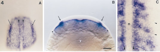
Fig. 4 Expression pattern of snail2 at the end of gastrulation and beginning of somitogenesis. Embryos were dissected from the yolk in A and C. (A) Embryo with three somites in dorsal view, anterior to the top. Two rows of strongly labeled snail2-expressing cells, called cephalic adaxial cells (arrowheads), are continuous with adaxial cells in the trunk bordering the unlabeled notochord. The cephalic adaxial cells define the border of the prechordal plate. Laterally, presumptive neural crest cells (arrows) begin to accumulate snail2 transcript. (B) An optical section in the presumptive head of an embryo at the same stage showing the mesendoderm (arrowheads), a very thin layer of cells expressing snail2, lying adjacent to the yolk cell (y). Medial to the lateral borders of the stained cephalic mesoderm, snail2 transcripts are revealed in presumptive neural crest cells (arrows). (C) Dorsal view of the trunk of a seven-somite embryo. The notochord (n) no longer contains snail2 transcript. Adaxial cells (large arrowheads) are strongly labeled as well as cells of the posterior part of the somites. Somitic furrows are indicated by small arrowheads. Scale bars, 25 μm (A), 40 μm (B), and 100 μm (C).
Reprinted from Developmental Biology, 172, Thisse, C., Thisse, B., and Postlethwait, J.H., Expression of snail2, a second member of the zebrafish Snail family, in cephalic mesendoderm and presumptive neural crest of wild-type and spadetail mutant embryos, 86-99, Copyright (1995) with permission from Elsevier. Full text @ Dev. Biol.