
|
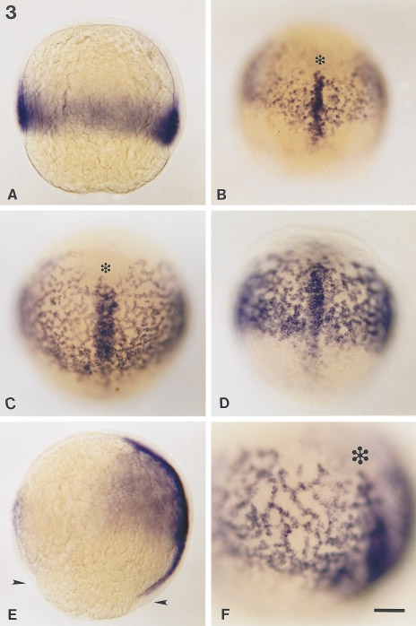
Fig. 3 Distribution of snail2 RNA during gastrulation. Transcripts from the snail2 gene were revealed by whole-mount in situ hybridization. (A) Optical section of an embryo at 65% epiboly oriented with its animal pole up. snail2 RNA appears in involuted mesendoderm. (B) Dorsal view of an embryo at 70% epiboly. The dorsal axial mesoderm appears as a nearly continuous strip of snail2 labeling about 3 cells wide coursing through the network of snail2-expressing cells. Presumptive hatching gland cells (asterisk) that form the anterior part of the prechordal plate do not express snail2. (C) Embryo at 80% epiboly in a dorsal–anterior view or (D) in dorsal–posterior view. The dorsal axis staining has become broader and extends beyond the stained web. (E) Optical section of an embryo at 90% epiboly. Dorsal, right, and animal pole, top. The axial snail2 expression domain extends posteriorly in the notochordal territory and reaches the margin (indicated by arrowheads). (F) High magnification of the left side of an embryo at 90% epiboly in the cephalic region. Cells labeled with snail2 probe form branching rows from 10 to 20 cells long. Scale bars, 25 μm (A–E); 40 μm (F).
Reprinted from Developmental Biology, 172, Thisse, C., Thisse, B., and Postlethwait, J.H., Expression of snail2, a second member of the zebrafish Snail family, in cephalic mesendoderm and presumptive neural crest of wild-type and spadetail mutant embryos, 86-99, Copyright (1995) with permission from Elsevier. Full text @ Dev. Biol.