
|
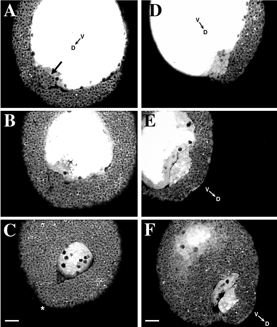
Fig. 4 The forerunner cell cluster does not involute during late epiboly. A-C and D-F show time-lapse recordings of two different embryos. The recordings are at slightly different focal planes and orientations. Scale bars, 50 μm. The embryo in D-F has a larger number of forerunner cells than the embryo in A-C. (A-C) A time-lapse series of an embryo stained with BODIPY 505/515. Vegetal pole view. The embryo in A-C is filmed with the blastopore facing down against the microscope coverslip. A wedge-shaped group of NEM/forerunner cells is visible at the leading edge of the blastoderm from 70%- to 90%-epiboly (black arrow). The yolk cell is brightly labeled with BODIPY 505/515. (A) The embryo is at 70%-epiboly. (B) At 90%-epiboly, the fore-runner cell cluster becomes depressed into a more ventral position as the germ ring shrinks in circumference. The forerunner cells separate from overlying EVL-NEM cells at this point in development. (C) The dorsal apex of the forerunner cell cluster is aligned with the presumptive tailbud, which is seen as a dorsal thickening (asterisk) in the germ ring. (D-F) The embryo of this time-lapse series was costained with BODIPY 505/515 (to label all the embryo’s blastomeres) and SYTO-11 (to enhance the visibility of the forerunner cluster). The time-lapse recording begins with an oblique view of the embryo’s dorsal side. This embryo was not immobilized in an agarose-ERM gel. During the recording, the embryo rolled into a position with its vegetal pole facing the coverslip (see E). The embryo was recentered in F. (D) Oblique view of the embryo at 70%-epiboly. The forerunner cell cluster, with more brightly labeled cells, is located at the leading margin of the blastoderm. (E) Vegetal pole view at 90%-epiboly. The forerunner cell cluster forms a bilaterally symmetric wedge at the distal margin of the advancing blastoderm. Dark nuclei are visible within the YSL. (F) At blastopore closure, the forerunner cell cluster forms the posterior limit of the embryonic axis. The embryo was followed until the tailbud stage when it was reimaged from a side view (see Fig. 5A).
Reprinted from Developmental Biology, 180(1), Cooper, M.S. and D'Amico, L.A., A cluster of noninvoluting endocytic cells at the margin of the zebrafish blastoderm marks the site of embryonic shield formation, 184-198, Copyright (1996) with permission from Elsevier. Full text @ Dev. Biol.