
|
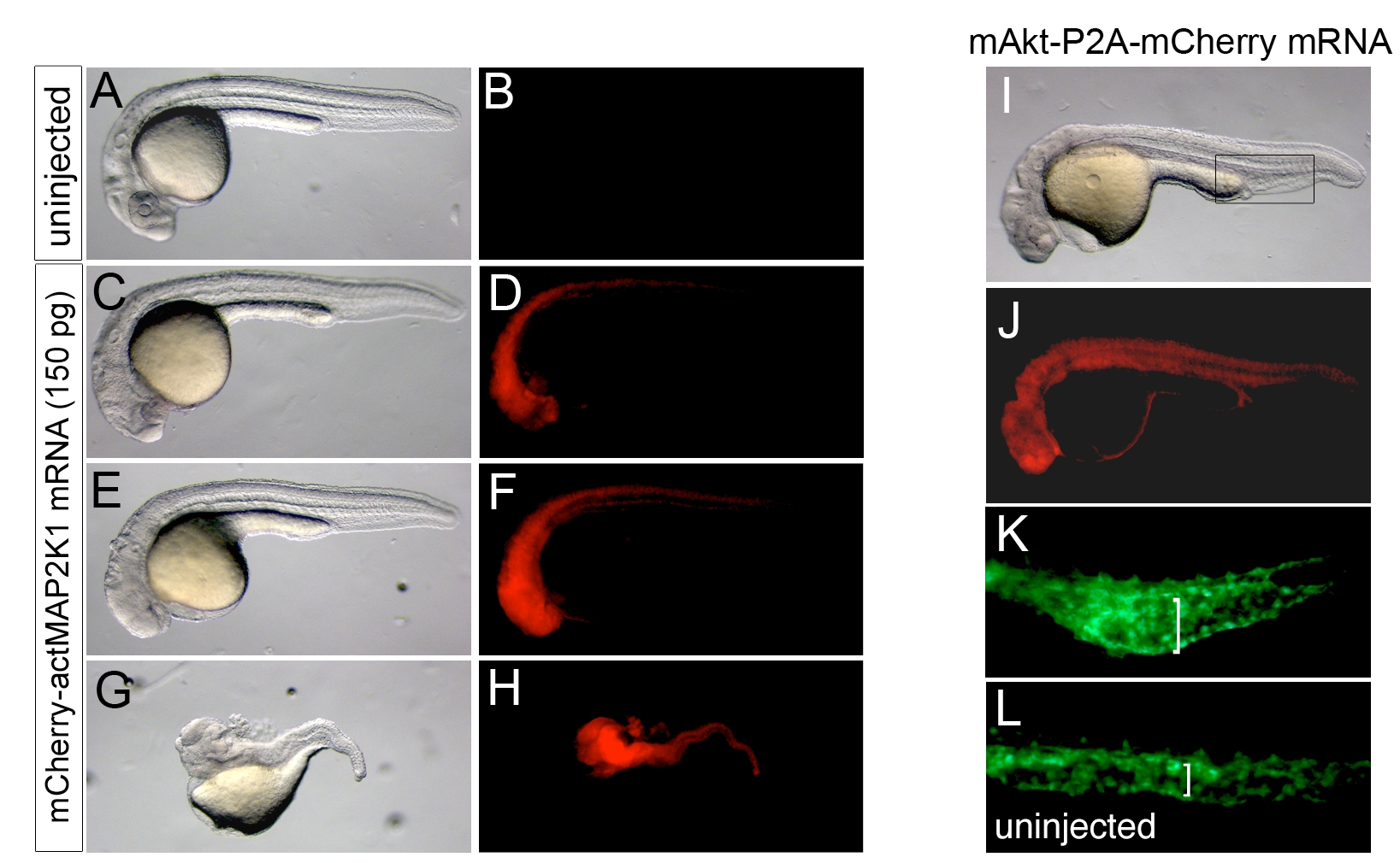
Fig. S2 Developmental defects in embryos with constitutive MAP2K1 or Akt activation. A, C, E, G, I – Transmitted light images; lateral views, anterior to the left, dorsal is up. B, D, F, H, J-L – Epifluorescent images. A, B. Uninjected wild type embryo, showing no red fluorescence. C-H. Embryos injected with 150 pg of mRNA encoding mCher-actMAP2K1 showing defects increasing in severity from C. slight cyclopia and reduction in eye size, E. complete absence of eyes and reduction in forebrain, to G. complete loss of trunk and head structures. In each case, the mCheractMAP2K1 fusion could be visualized by red fluorescence (D, F, H). I-K. Embryo injected with 200 pg of mRNA encoding mAkt-2A-mCherry. I. At 27 hours post fertilization, injected embryos displayed slight pericardial edema and smaller eyes. J. Red fluorescence was also visible at 30 hpf. K. Tg(fli1:egfp)y1 embryos injected with mAkt-2A-mCherry mRNA also displayed enlarged caudal vein lumens (bracket) compared to L. uninjected Tg(fli1:egfp)y1 siblings. The presence of pericardial edema, small eyes, and expanded caudal vein suggests that early activation of Akt may lead to mild ventralization of the zebrafish embryo.
Reprinted from Developmental Biology, 329(2), Covassin, L.D., Siekmann, A.F., Kacergis, M.C., Laver, E., Moore, J.C., Villefranc, J.A., Weinstein, B.M., and Lawson, N.D., A genetic screen for vascular mutants in zebrafish reveals dynamic roles for Vegf/Plcg1 signaling during artery development, 212-226, Copyright (2009) with permission from Elsevier. Full text @ Dev. Biol.