
|
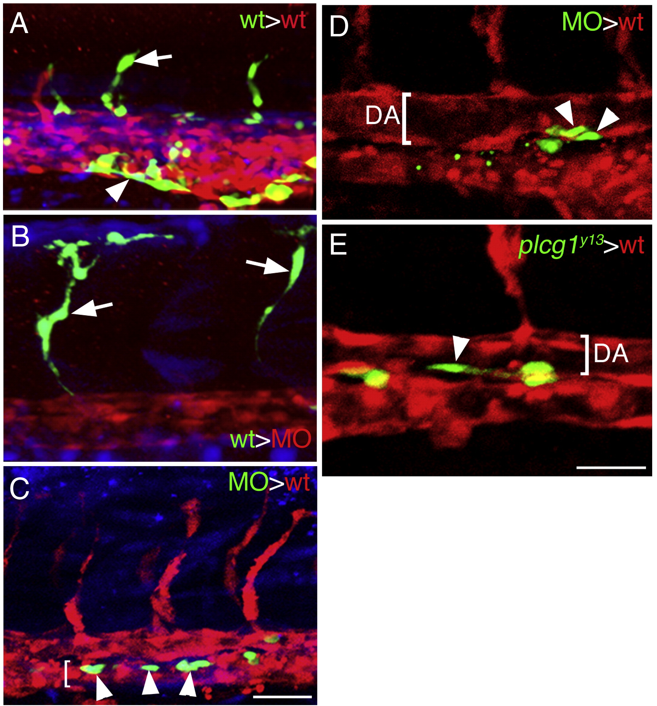
Fig. 7 Plcg1 is required autonomously in endothelial cells for segmental artery sprouting. (A.–E) Confocal fluorescent micrographs of trunk blood vessels at approximately 27 hpf. Lateral views, anterior to the left, dorsal is up. In all images, host cells are expressing dsRedEx (red), while donor cells are expressing enhanced green fluorescent protein (green); in (A–C) donor cells are also labeled with Cascade Blue (blue). (A) Control embryo derived from transplantation of wild type donor Tg(fli1a:egfp)y1 cells into a Tg(fli1ep:dsredex)um13 host. Donor contribution to a developing segmental artery (white arrow) and the caudal vein (white arrowhead) is indicated. (B) Contribution of donor Tg(fli1a:egfp)y1 cells to a segmental artery (white arrow) in a plcg1 MO injected Tg(fli1ep:dsredex)um13 host. (C) Donor plcg1-MO injected Tg(fli1a:egfp)y1 cells (white arrows) within the posterior cardinal vein (lumen indicated by a white bracket) in a wild type Tg(fli1ep:dsredex)um13 host. (D) Donor plcg1-MO injected Tg(fli1a:egfp)y1 cells (white arrowheads) within the ventral wall of the dorsal aorta (DA, lumen denoted by a white bracket) in a wild type Tg(fli1ep:dsredex)um13 host. (E) Donor Tg(fli1a:egfp)y1;plcg1y13 mutant cells (white arrowheads) within the ventral wall of the dorsal aorta (DA, lumen denoted by a white bracket) in a wild type Tg(fli1ep:dsredex)um13 host. (A–C) Scale bar is 50 μM. (D, E) Scale bar is 30 μM.
Reprinted from Developmental Biology, 329(2), Covassin, L.D., Siekmann, A.F., Kacergis, M.C., Laver, E., Moore, J.C., Villefranc, J.A., Weinstein, B.M., and Lawson, N.D., A genetic screen for vascular mutants in zebrafish reveals dynamic roles for Vegf/Plcg1 signaling during artery development, 212-226, Copyright (2009) with permission from Elsevier. Full text @ Dev. Biol.