
|
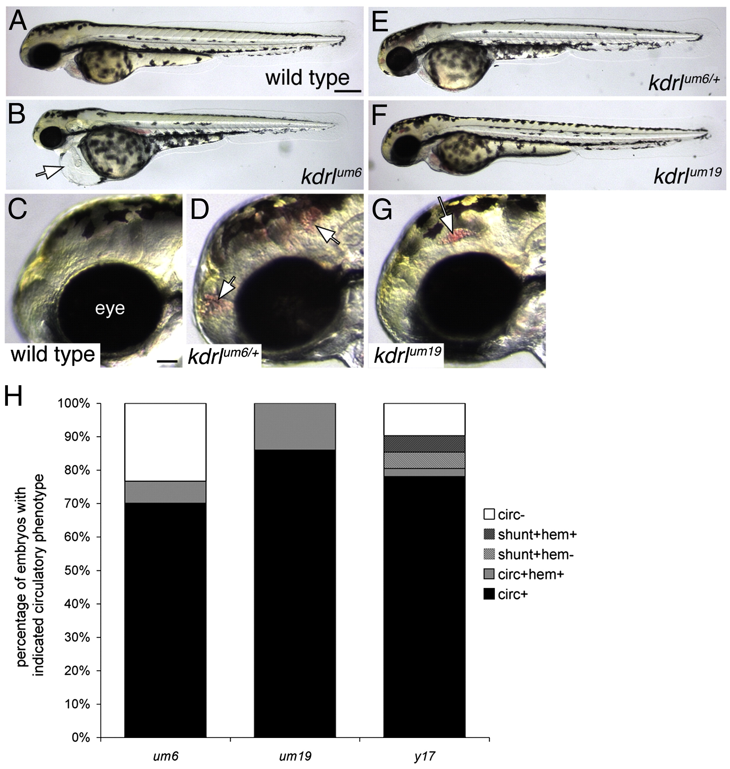
Fig. 3 Defects in circulatory function in kdrl mutants. (A–G) Transmitted light images of wild type and kdrl mutant zebrafish embryos at 55 h post fertilization. Lateral views, anterior to the left, dorsal is up. (A) Wild type sibling embryo. Scale bar is 250 μM. (B) kdrlum6 mutant embryo displaying cardiac edema (white arrow). (C) Eye (indicated) and forebrain region of wild type sibling embryo. Scale bar is 50 μM. (D) Cranial hemorrhage (white arrows) in a heterozygous carrier of the kdrlum6 allele. (E) kdrlum6 heterozygous embryo. (F) kdrlum19 mutant embryo. (G) Eye and forebrain region of a kdrlum19 mutant embryo. A cranial hemorrhage is indicated by the white arrow. (H) Quantification of circulatory defects in clutches of embryos derived from incrosses of um6, um19, and y17 heterozygous carriers; circ — circulation, shunt — abnormal circulatory connection between dorsal aorta and posterior cardinal vein, hem — cranial hemorrhage.
Reprinted from Developmental Biology, 329(2), Covassin, L.D., Siekmann, A.F., Kacergis, M.C., Laver, E., Moore, J.C., Villefranc, J.A., Weinstein, B.M., and Lawson, N.D., A genetic screen for vascular mutants in zebrafish reveals dynamic roles for Vegf/Plcg1 signaling during artery development, 212-226, Copyright (2009) with permission from Elsevier. Full text @ Dev. Biol.