
|
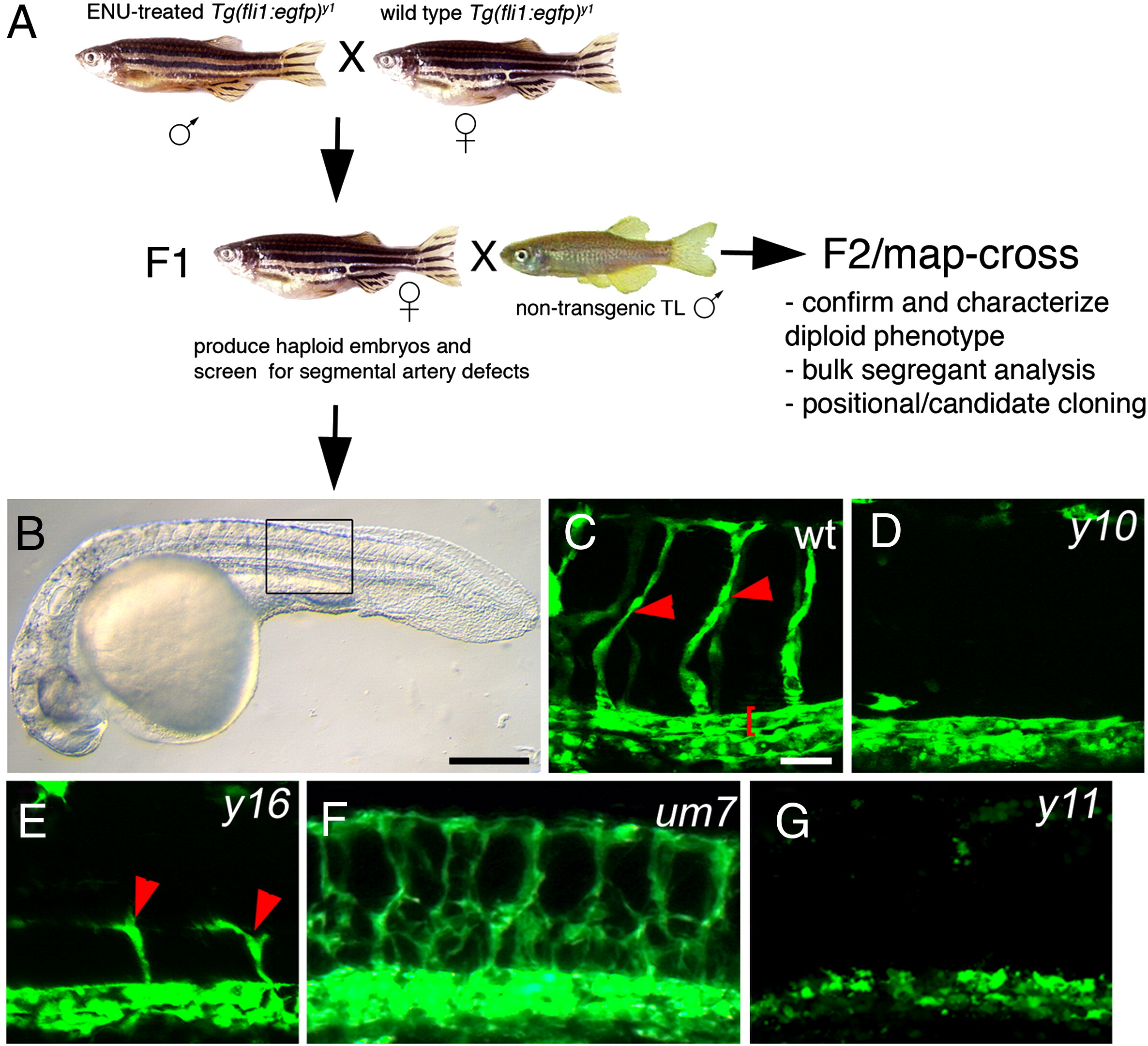
Fig. 1 A transgenic haploid screen for vascular mutants. (A) Breeding strategy for transgenic haploid screen. (B) Transmitted light image of a Tg(fli1a:egfp)y1 haploid embryo. (C) Segmental arteries (red arrowheads) in a wild type Tg(fli1a:egfp)y1 haploid embryo. Red bracket denotes dorsal aorta. (D) Loss of segmental arteries in a y10 mutant haploid embryo. (E) Partial segmental artery sprouts (red arrowheads) in a y16 mutant haploid embryo. (F) Excessive segmental artery branching in a um7 mutant haploid embryo. (G) Loss of segmental artery formation and failure to form the dorsal aorta in a y11 haploid mutant embryo. (B–G) Lateral views, anterior to the left, dorsal is up. (C–E, G). Confocal fluorescent micrographs. (F) Epifluorescent image. (B) Scale bar is 250 μM. (C–G) Scale bar is 50 μM.
Reprinted from Developmental Biology, 329(2), Covassin, L.D., Siekmann, A.F., Kacergis, M.C., Laver, E., Moore, J.C., Villefranc, J.A., Weinstein, B.M., and Lawson, N.D., A genetic screen for vascular mutants in zebrafish reveals dynamic roles for Vegf/Plcg1 signaling during artery development, 212-226, Copyright (2009) with permission from Elsevier. Full text @ Dev. Biol.