Image
Figure Caption
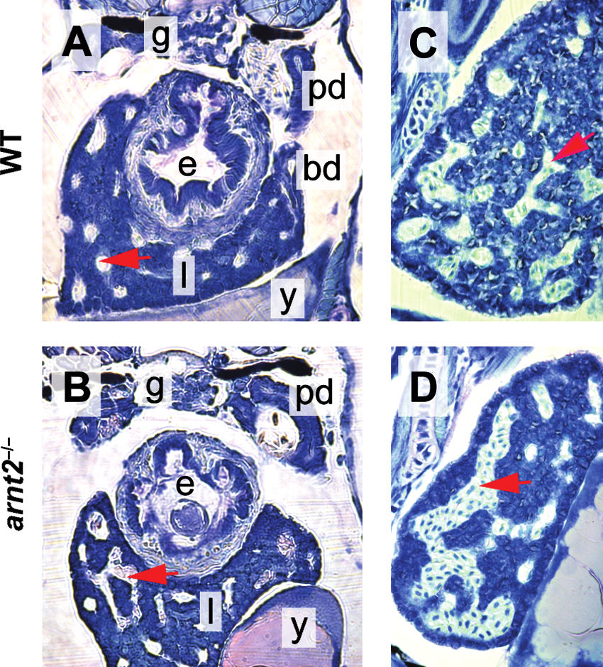
Fig. 9 Altered liver sinusoid morphology in arnt2-/- larvae at 120 hpf. Photomicrographs of sections of the liver of a representative WT larva (A, C) and arnt2-/- mutant (B, D) are shown. Compared to WT, the arnt2-/- liver is characterized by enlarged sinusoids engorged with blood that have merged to form large labyrinth-like vascular channels. A red arrowhead points to a blood-filled sinusoid in each of the sections. e, Esophagus; l, liver; y, yolk; g, glomerulus; pd, pronephric duct; bd, bile duct.
Figure Data
Acknowledgments
This image is the copyrighted work of the attributed author or publisher, and
ZFIN has permission only to display this image to its users.
Additional permissions should be obtained from the applicable author or publisher of the image.
Full text @ Zebrafish

