
|
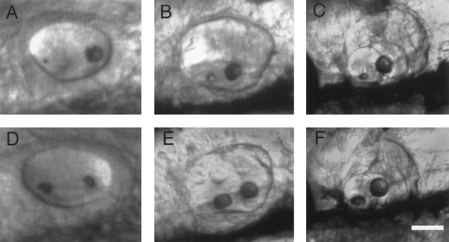
Fig. 6 Otolith development following modification. (A–C) Development of the same wild-type ear depicted in Figs. 5B–5F following enhancement of the posterior otolith with laser tweezers. (A) At 24 h, approximately 2 h after laser treatment, the posterior otolith is still many times the size of the anterior otolith. At 2 (B) and 6 days (C), the posterior otolith is nearly normal in size, but the anterior otolith remains much smaller than normal. (D–F) Development of a wild-type control embryo at 24 h (D), 2 days (E), and 6 days (F). Anterior is to the left and dorsal is upward in all panels. Scale bar, 25 (A and D), 45 (B and E), and 100 μm (C and F).
Reprinted from Developmental Biology, 191(2), Riley, B.B., Zhu, C., Janetopoulos, C., and Aufderheide, K.J., A critical period of ear development controlled by distinct populations of ciliated cells in the zebrafish, 191-201, Copyright (1997) with permission from Elsevier. Full text @ Dev. Biol.