
Image
|
Figure Caption
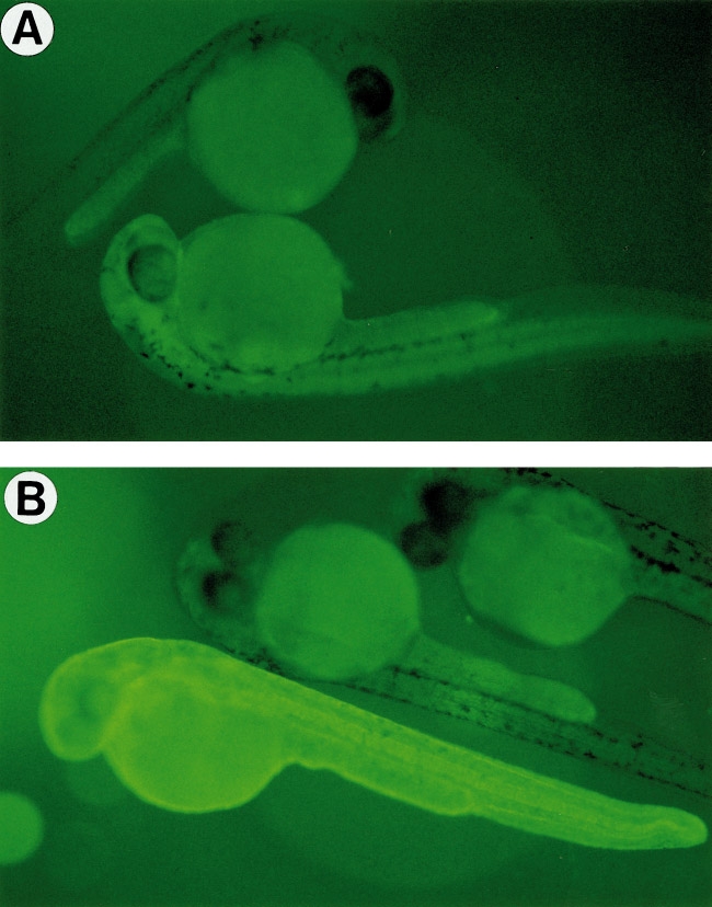
Fig. 6 Expression of GFP in transgenic embryos generated using β-actin–GFP. Chorions were removed. Nonfluorescent embryos are nontransgenic siblings. (A) A typical transgenic embryo at 28 h. (B) A transgenic embryo from the most fluorescent line at 28 h.
Acknowledgments
This image is the copyrighted work of the attributed author or publisher, and
ZFIN has permission only to display this image to its users.
Additional permissions should be obtained from the applicable author or publisher of the image.
Reprinted from Developmental Biology, 192, Higashijima, S., Okamoto, H., Ueno, N., Hotta, Y., and Eguchi, G., High-frequency generation of transgenic zebrafish which reliably express GFP in whole muscles or the whole body by using promoters of zebrafish origin, 289-299, Copyright (1997) with permission from Elsevier. Full text @ Dev. Biol.