
Image
|
Figure Caption
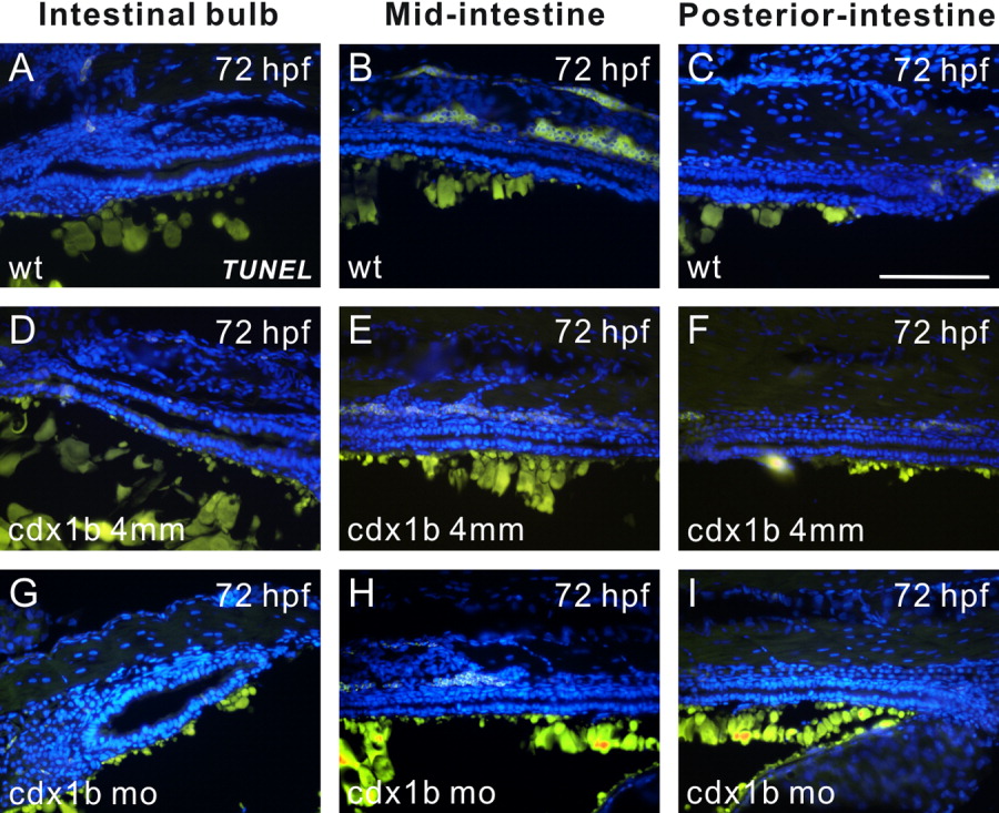
Fig. 10 TUNEL assay revealed a low level of apoptosis in cdx1b morphants. A-I: Paraffin sagittal sections corresponding to the intestinal bulb (A, D, G), mid-intestine (B, E, H), and posterior-intestine (C, F, I) of respective 72-hr post-fertilization (hpf) wild type (A-C), cdx1b-4mm-MO-injected (D-F), and cdx1b MO-injected (G-I) embryos treated with TUNEL reactions using FITC-dUTP (green). Nuclei were stained with DAPI (blue). Embryos in A-C and G-I are oriented with the anterior to the left, while those in D-F are oriented with the anterior to the right. Scale bars = 100 μm.
Figure Data
Acknowledgments
This image is the copyrighted work of the attributed author or publisher, and
ZFIN has permission only to display this image to its users.
Additional permissions should be obtained from the applicable author or publisher of the image.
Full text @ Dev. Dyn.