
|
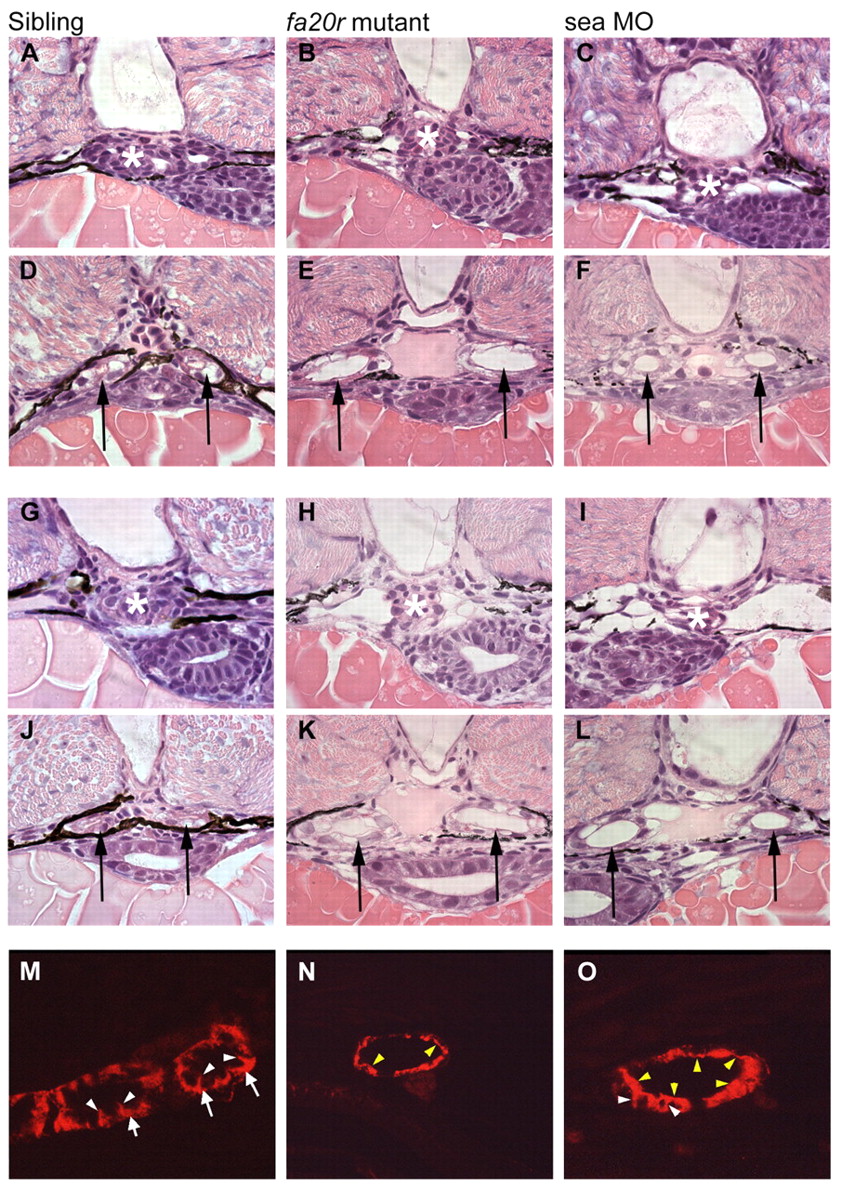
Fig. 5 Pronephric cyst phenotypes in seahorse mutants and sea MO-injected embryos. (A-F) At 2 dpf, the glomerular region (asterisks) appears normal in siblings (A) and seafa20r mutants (B), and slightly dilated in sea MO-injected embryos (C). In the same embryos, the medial tubules (arrows) were significantly dilated both in seafa20r mutants (E) and sea MO-injected embryos (F), compared with siblings (D). (G-L) At 2.5 dpf, the glomeruli (asterisks) in both the seafa20r mutants (H) and sea MO-injected embryos (I) were dilated compared with siblings (G). These same embryos have dilations in the medial tubules (arrows) in seafa20r mutants (K) and sea MO-injected embryos (L) compared with siblings (J). (M-O) At 3 dpf, siblings (M) display basolateral localization of Na+/K+-ATPase (red) in the pronephric epithelium (arrows; white arrowheads indicate lateral staining), whereas the sea mutants (N,O) display altered localization of Na+/K+-ATPase diffusely at apical (yellow arrowheads) and lateral membranes (white arrowheads).