
|
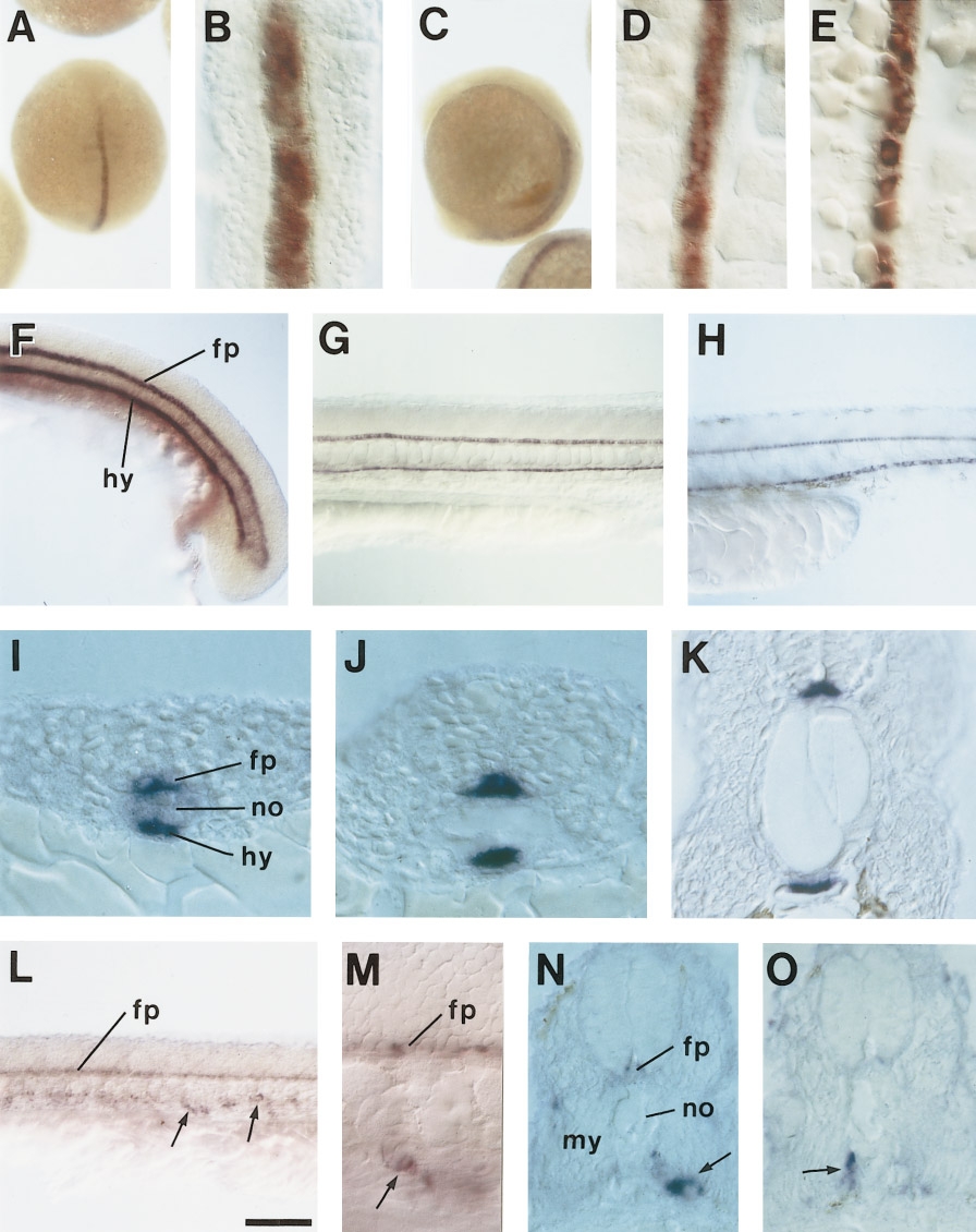
Fig. 4 (A–K) mindin1 and (L–O) mindin2 mRNA expression in developing embryos. (A and B) mindin1 expression in 11-h embryos. Dorsal view. A higher magnification view of a dissected embryo is presented in B. Expression of mindin1 first appears in the axial mesodermal region. (C) Lateral view of an embryo at 13 h. (D) A dorsal view of a 14-h embryo in higher magnification. The focus is just dorsal to the notochord. A continuous row of cells, which are precursors of the floor plate cells, are labeled. (E) The same embryo shown in D. The focus is just ventral to the notochord. Noncontiguous cells, which are precursors of the hypochordal cells, are labeled. (F–H) Lateral views of 17- (F), 24- (G), and 33-h (H) embryos. Two contiguous rows of cells just dorsal or ventral to the notochord are labeled. The former is the floor plate and the latter is the hypochord. (I–K) Cross sections of 12- (I), 17- (J), and 30-h (K) embryos. In all developmental stages, mindin1 is expressed in the floor plate and the hypochord, either of which consists of a single row of cells. In the early stage (I), mindin1 is also expressed weakly in the notochord. (L and M) mindin2 expression in a 26-h embryo. Lateral view. A higher magnification view of the same embryo shown in L is presented in M. mindin2 is expressed in the floor plate cells. mindin2 is also expressed in the mesenchymal cells in the ventral region of the embryo (arrows). (N and O) Cross section of 32-h embryos. mindin2-positive mesenchymal cells are located between the ventral region of the myotome and endodermal cells (arrow in N) or between the ventral region of the notochord and the myotome (arrow in O). These cells are likely to be sclerotomal cells. fp, floor plate; no, notochord; hy, hypochord; my, myotome. Scale bar, 270 μm in A and C; 44 μm in B, D, and E; 100 μm in F–H and L; 17 μm in I–K, N, and O; and 33 μm in M.
Reprinted from Developmental Biology, 192, Higashijima, S., Nose, A., Eguchi, G., Hotta, Y., and Okamoto, H., Mindin/F-spondin family: novel ECM proteins expressed in the zebrafish embryonic axis, 211-227, Copyright (1997) with permission from Elsevier. Full text @ Dev. Biol.