
|
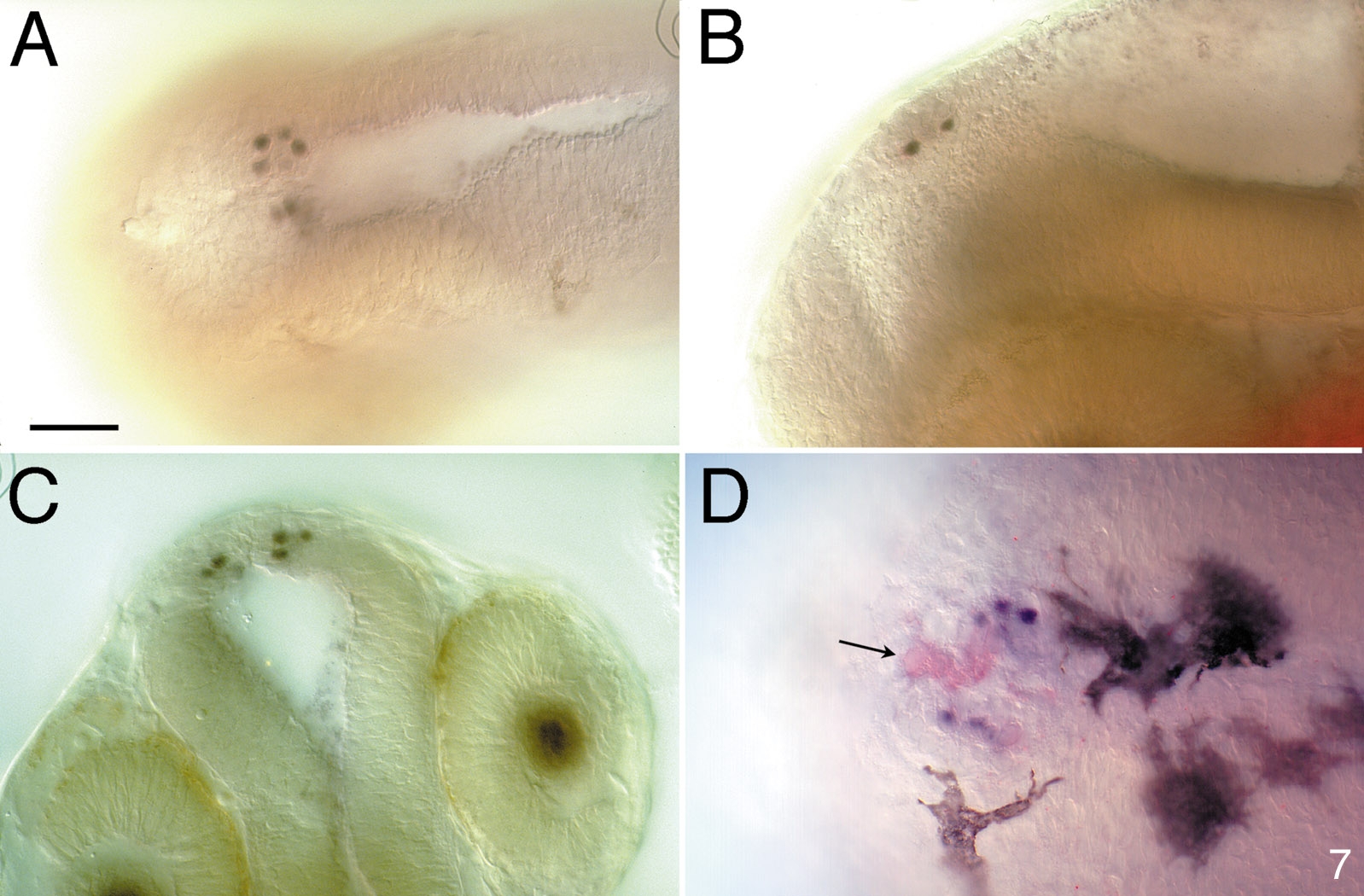
Fig. 7 Expression of Lim3 in the epiphysis is restricted to a subset of projection neurons. (A–C) Four to five Lim3-expressing cells are seen on each side of the midline in a ventrolateral position in the epiphysis of a 28-h embryo. (A) Dorsal view. (B) Lateral view. (C) Anterior view. This optical section through the epiphysis, with the diencephalon in focus, is slightly deeper than in Fig. 5D. (D) Dorsal view of a 2-day-old embryo. Whole-mount embryos were double labeled with anti-S-antigen (red) and anti-Lim3 (blue, nuclei). Four to five projection neurons express Lim3 protein. Several cells, especially in the center of the epiphysis, express the photoreceptor marker, S-antigen (arrow). By this stage several prominent melanocytes are seen. Scale bar is 50 μm.
Reprinted from Developmental Biology, 192, Glasgow, E., Karavanov, A.A., and Dawid, I.B., Neuronal and neuroendocrine expression of lim3, a LIM class homeobox gene, is altered in mutant zebrafish with axial signaling defects, 405-419, Copyright (1997) with permission from Elsevier. Full text @ Dev. Biol.