
Image
|
Figure Caption
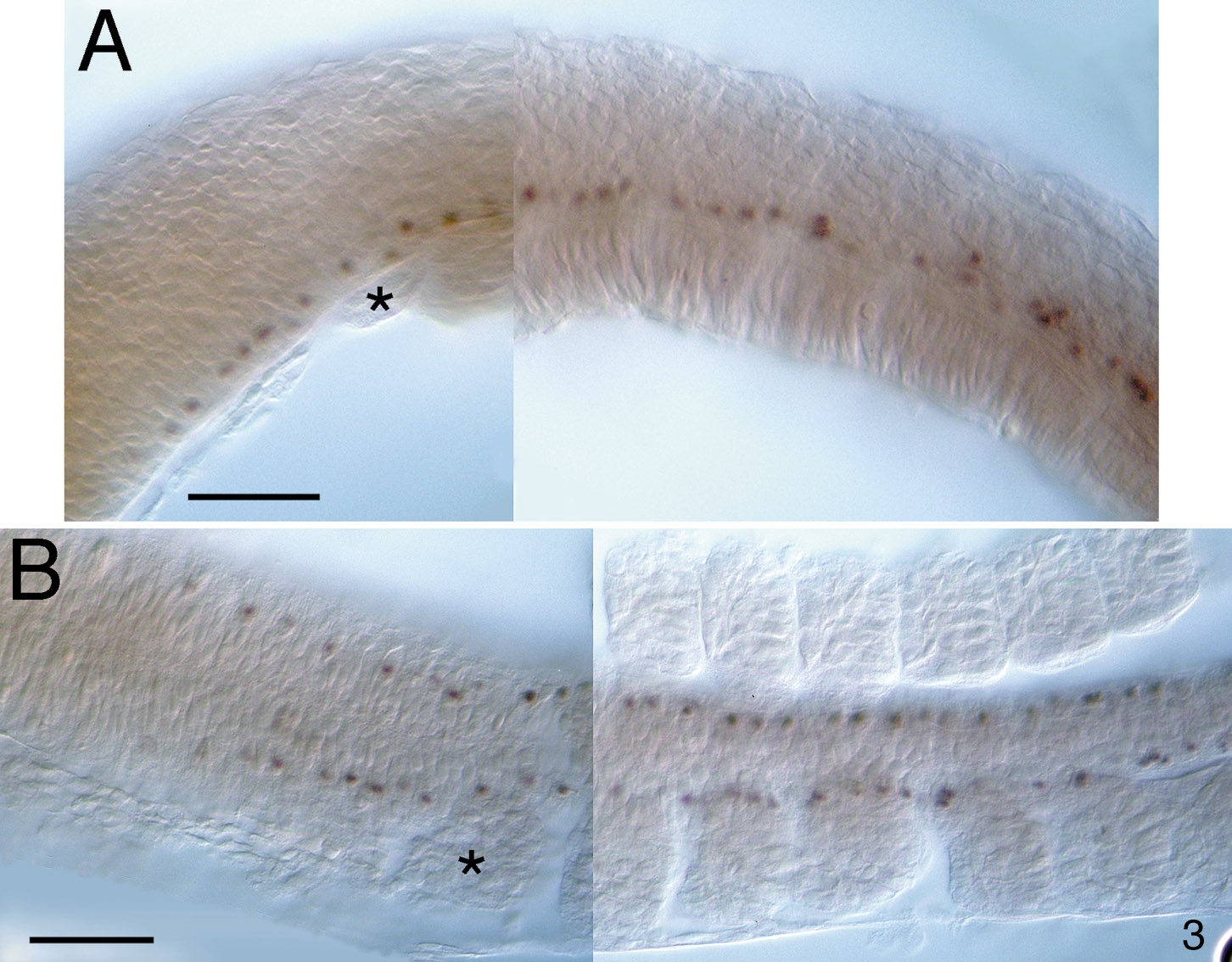
Fig. 3 Expression of Lim3 protein in 10-somite-stage zebrafish embryos. (A and B) Ten-somite-stage embryos stained as whole mounts were dissected from the yolk and flatted under a coverslip. Anterior is to the left. Three to four Lim3-positive cells are seen in each segment associated with each somite. The first somite is indicated by an asterisk. Scale bars are 50 μm. (A) Lateral view, dorsal is up. Lim3-positive cells are in a ventral position in the hindbrain and spinal cord. (B) Dorsal view. The Lim3-positive cells in the hindbrain appear to be continuous with those of the spinal cord.
Figure Data
Acknowledgments
This image is the copyrighted work of the attributed author or publisher, and
ZFIN has permission only to display this image to its users.
Additional permissions should be obtained from the applicable author or publisher of the image.
Reprinted from Developmental Biology, 192, Glasgow, E., Karavanov, A.A., and Dawid, I.B., Neuronal and neuroendocrine expression of lim3, a LIM class homeobox gene, is altered in mutant zebrafish with axial signaling defects, 405-419, Copyright (1997) with permission from Elsevier. Full text @ Dev. Biol.