
|
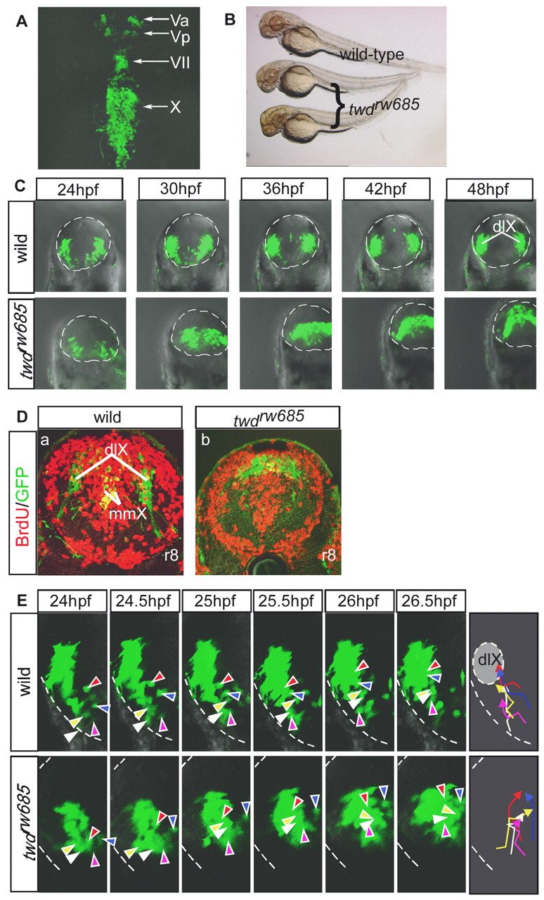
Fig. 3 The twdrw685 mutant shows aberrant migration of vagus motor neuron progenitors. (A) Dorsal view of the twdrw685 embryos at 50 hpf (rostral towards the top). (B) Morphology of wild-type and twdrw685 embryos at 2 dpf. (C) Time-lapse imaging of the behavior of the vagus motor neuron progenitors in wild-type (upper row) and twdrw685 (lower row) embryos. The broken line outlines the hindbrain. dlX, dorsolateral motor nucleus of the vagus. (D) Vagus motor neurons and incorporated BrdU at 38 hpf were detected by anti-BrdU (red) and anti-GFP (green) antibodies in wild-type (a) and twdrw685 (b) embryos at 72 hpf (cross-sections; dorsal towards the top). mmX, medial motor nucleus of the vagus. (E) Migratory pathways of five randomly chosen progenitors of the dlX, indicated by different-colored arrowheads. The broken lines show the outline of neural tubes. The lines indicate the trajectories of the individual neurons and correspond to the colors shown in the right-most schematic diagrams.