
Image
|
Figure Caption
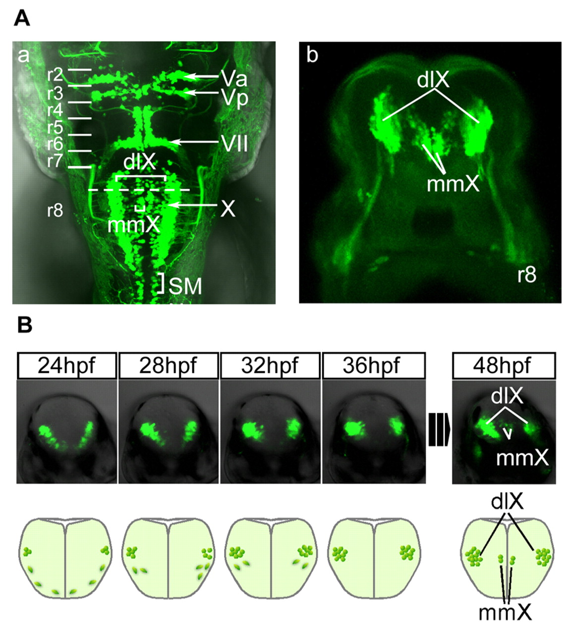
Fig. 2 Visualization and live imaging of vagus motor nuclei in isl1:GFP transgenic zebrafish. (A) Fluorescent images of the hindbrain of the isl1:GFP transgenic zebrafish. (a) Dorsal view (rostral towards the top) and (b) cross-section (dorsal towards the top) at the broken line shown in a. (B) Time-lapse imaging of the development of the vagus motor nuclei (upper row), accompanied by the schematic diagrams (lower row; dorsal towards the top). dlX, dorsolateral motor nucleus of the vagus; mmX, medial motor nucleus of the vagus; SM, spinal motor neurons.
Figure Data
Acknowledgments
This image is the copyrighted work of the attributed author or publisher, and
ZFIN has permission only to display this image to its users.
Additional permissions should be obtained from the applicable author or publisher of the image.
Full text @ Development