
|
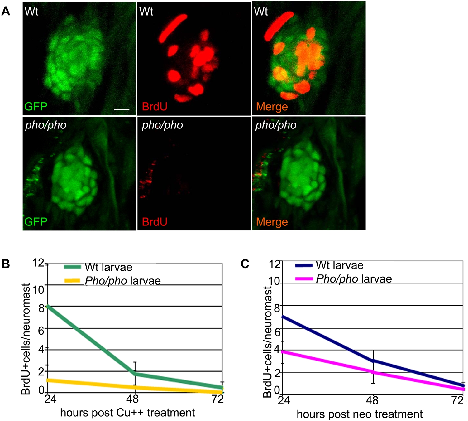
Fig. 5 Supporting cells do not divide as efficiently after hair cell damage in the phoenix mutant neuromast.
(A) BrdU staining (red, in the middle and right panels) in the SCM1::GFP transgenic wild-type (top) and the SCM1::GFP transgenic phoenix mutant neuromast (bottom) of recovering larvae (n = 20/20), 24h after copper treatment. The supporting cells are counterstained with GFP antibodies (green in left and right panels). (B) Quantification of the BrdU positive cells/neuromast in wild-type (green line) and mutant (yellow line) recovering neuromasts, at +24h (n = 11/10), +48h (n = 10/6), and +72h (n = 7/6) after copper treatment. Larvae are exposed to BrdU for 6h prior to being sacrificed. (C) Quantification of the BrdU positive cells/neuromast in wild-type (blue line) and mutant (pink line) recovering larvae, at +24h (n = 22/40), +48h (n = 20/33), and +72h (n = 13/32) after neomycin exposure. – 10 microns in (A).