
Image
|
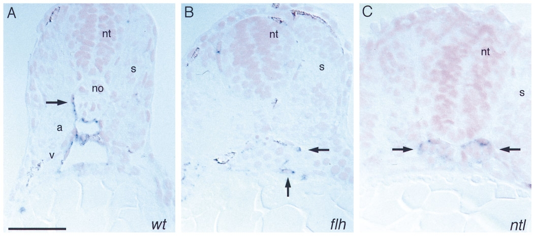
Figure Caption
Fig. 7 Histological analysis of whole mount RNA in situ hybridization with flk performed on 30 somite stage embryos (24 hpf). (A) Wild type, (B) floating head, (C) no tail. (A) The wild-type embryo clearly shows the two axial vessels, dorsal aorta (a) and axial vein (v) and a sprout of an intersomitic vessel (arrow). (B, C) No axial vessels can be observed in floating head or no tail. Arrows point to flk-expressing cells. Sections were counterstained with Nuclear Fast Red. Dorsal is up. Bar, 50 μm.
Acknowledgments
This image is the copyrighted work of the attributed author or publisher, and
ZFIN has permission only to display this image to its users.
Additional permissions should be obtained from the applicable author or publisher of the image.
Reprinted from Developmental Biology, 183(1), Fouquet, B., Weinstein, B.M., Serluca, F.C., and Fishman, M.C., Vessel patterning in the embryo of the zebrafish: guidance by notochord, 37-48, Copyright (1997) with permission from Elsevier. Full text @ Dev. Biol.