
|
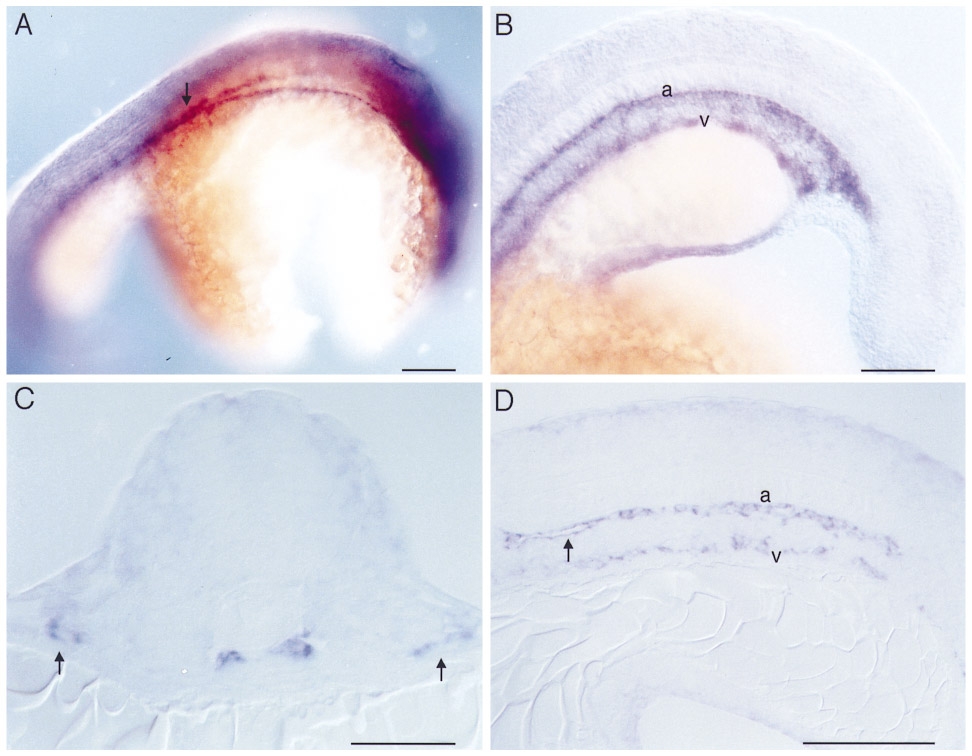
Fig. 4 Whole mount RNA in situ hybridization with flk on 24 somite stage embryos (21 hpf). (A) Dorsolateral view showing the paired dorsal aortae in the area of the hindbrain and their junction in the anterior trunk (arrow). (B) Lateral view of the posterior trunk and tail showing the primordia of the dorsal aorta (a) and the axial vein (v). (C) Transverse section through the anterior trunk at the level of the first somite. The two primordia of the dorsal aortae lie just beneath the notochord. Laterally located (arrows) are precursors of the bilateral ducti of Cuvier. (D) Sagittal section of the posterior trunk showing the primordia of the dorsal aorta (a) and the ventral vein (v). Arrow indicates beginning lumen formation in the dorsal aorta. No intersomitic vessels are yet formed. Dorsal is up, and anterior is to the right in (A) and to the left in (B, D). Bars, 100 μm (A, B), 50 μm (C, D).
Reprinted from Developmental Biology, 183(1), Fouquet, B., Weinstein, B.M., Serluca, F.C., and Fishman, M.C., Vessel patterning in the embryo of the zebrafish: guidance by notochord, 37-48, Copyright (1997) with permission from Elsevier. Full text @ Dev. Biol.