
|
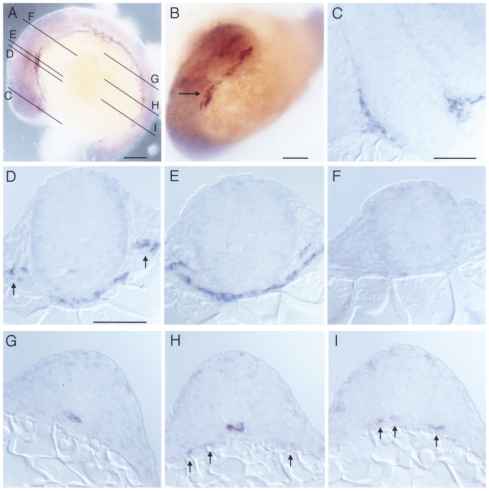
Fig. 3 Whole mount RNA in situ hybridization with flk performed on 14 somite stage embryos (16 hpf). (A) Lateral view and (B) anterior view showing angioblastic areas in the embryo. Lines in (A) indicate planes of sectioning for panels C through I. Arrow in (B) indicates position of first aortic arch. (C) Section at the level of the forebrain and eye vesicles, showing angioblastic cells in the ventrally to the brain located plexus. (D) Section at the level of the endocardial primordium which has reached the midline by this stage. Arrows indicate the primordia of the internal carotid artery. (E) Section at the level of the aortic arch primordium. (F) Section at the level of the hindbrain, which lacks flk-positive cells at this stage. (G –I) Sections at various planes through the trunk. Angioblasts have reached the midline below the notochord (G, H) forming the primordium of the dorsal aorta. Other angioblasts are still located laterally close to the yolk (arrows). (I) In the posterior trunk angioblastic cells have not yet reached the midline (arrows). Dorsal is up (A, C–I) and anterior is to the left (A). Bars, 100 μm (A, B), 50 μm (C–I).
Reprinted from Developmental Biology, 183(1), Fouquet, B., Weinstein, B.M., Serluca, F.C., and Fishman, M.C., Vessel patterning in the embryo of the zebrafish: guidance by notochord, 37-48, Copyright (1997) with permission from Elsevier. Full text @ Dev. Biol.