
Image
|
Figure Caption
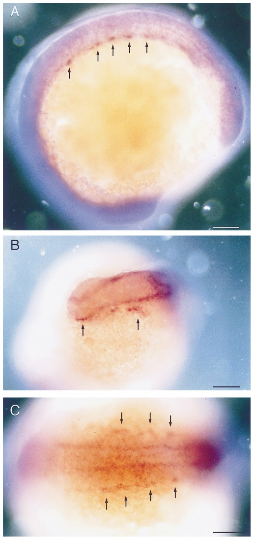
Fig. 2 Whole mount RNA in situ hybridization with flk performed on 12 somite stage embryos (15 hpf). (A) Lateral view and (C) dorsal view showing the bilateral angiogenic cell clusters in the trunk (arrows). (B) Anterior view showing the endocardial and cephalic angioblasts (arrows). Dorsal is up (A) and anterior is to the left (A). Bars, 100 μm.
Acknowledgments
This image is the copyrighted work of the attributed author or publisher, and
ZFIN has permission only to display this image to its users.
Additional permissions should be obtained from the applicable author or publisher of the image.
Reprinted from Developmental Biology, 183(1), Fouquet, B., Weinstein, B.M., Serluca, F.C., and Fishman, M.C., Vessel patterning in the embryo of the zebrafish: guidance by notochord, 37-48, Copyright (1997) with permission from Elsevier. Full text @ Dev. Biol.