
|
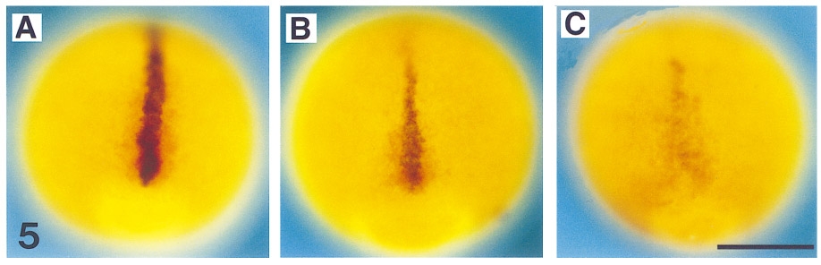
Fig. 5 shh expression in flh and cyc;flh mutants during late gastrulation. Dorsal views (anterior to the top) of whole-mount RNA in situ hybridization at 90% epiboly (9 hr) with antisense probes against shh in blue and myoD in red. At 90% epiboly shh is present in the midline of wild-type embryos (A). In flh mutants, shh is still present at the midline, but the expression levels are decreased compared to wild types (B). In cyc;flh mutants, the shh expression levels are dramatically decreased, but consistently present (C). myoD expression is in regions that will become paraxial mesoderm indicating that the shh expression corresponds to the trunk region of the embryo. Scale bar, 250 μm.
Reprinted from Developmental Biology, 187(2), Beattie, C.E., Hatta, K., Halpern, M.E., Liu, H., Eisen, J.S., and Kimmel, C.B., Temporal separation in the specification of primary and secondary motoneurons in zebrafish, 171-182, Copyright (1997) with permission from Elsevier. Full text @ Dev. Biol.