
|
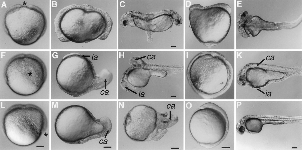
Fig. 7 Morphogenesis in janus-mutant embryos. The figure comprises the most common phenotypes observed. First three columns: Blastoderms are completely separated at the shield stage. Last two columns: Blastoderms are partially fused at the shield stage. (A –E) Shield is bisected among the two blastoderms. (F–K) Shield is restricted to one of the two blastoderms near the plane of division that bisects among them. (L–P) Shield is restricted to one of the two blastoderms opposite of the division plane that bisects among them. See text for detailed description of morphogenetic events. Lateral views. Asterisks indicate position of shield. ca, complete anterior axis; ia, incomplete anterior axis. Scale bars are 100 μm.
Reprinted from Developmental Biology, 184(1), Abdelilah, S. and Driever, W., Pattern formation in janus-mutant zebrafish embryos, 70-84, Copyright (1997) with permission from Elsevier. Full text @ Dev. Biol.