
Image
|
Figure Caption
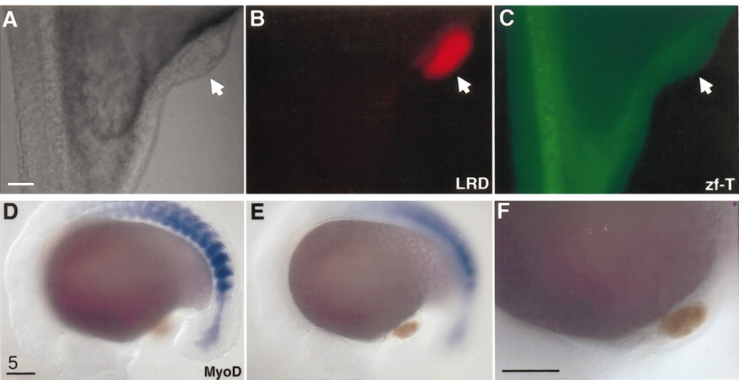
Fig. 5 Neither axial nor paraxial mesoderm is present within 80% epiboly hindbrain transplants. (A–C) Zf-T antibody detected host notochord cells (C), but failed to detect any cells within the graft (arrow in all panels). (D–F) In situ hybridization of MyoD (blue) labeled intensely the paraxial mesoderm (presumptive somites) of the host, but failed to give any signal within the graft (orange). All scale bars: 100 μm.
Acknowledgments
This image is the copyrighted work of the attributed author or publisher, and
ZFIN has permission only to display this image to its users.
Additional permissions should be obtained from the applicable author or publisher of the image.
Reprinted from Developmental Biology, 197, Woo, K. and Fraser, S.E., Specification of the hindbrain fate in the zebrafish, 283-296, Copyright (1998) with permission from Elsevier. Full text @ Dev. Biol.