
|
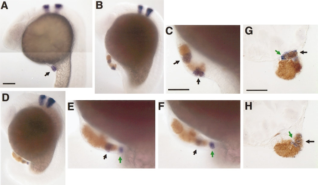
Fig. 6 Committed hindbrain cells express coherent patches or stripes of Krox20. (A) A single patch of Krox20 (arrow) is present in the 80HV graft. (B, C) Two distinct stripes of Krox20-positive cells (blue color) are expressed by the 80HV graft. Note that both Krox20 domains (C, arrows) are within the biotin-visualized lineage tracer territory (orange). (D–F) Another example with two distinct domains of Krox20: one stripe of Krox20 is expressed by grafted cells (black arrow); the other patch is expressed by host cells (devoid of lineage label that marks donor cells (green arrow). This host-derived Krox20 domain is continuous with the graft-derived structure extending outward from the yolk. (G, H) Histological sections of different embryos illustrating that graft-derived (black arrows) and host-derived (green arrows) Krox20-expressing cells are intermingled in the same structure. All scale bars: 100 μm.
Reprinted from Developmental Biology, 197, Woo, K. and Fraser, S.E., Specification of the hindbrain fate in the zebrafish, 283-296, Copyright (1998) with permission from Elsevier. Full text @ Dev. Biol.