
|
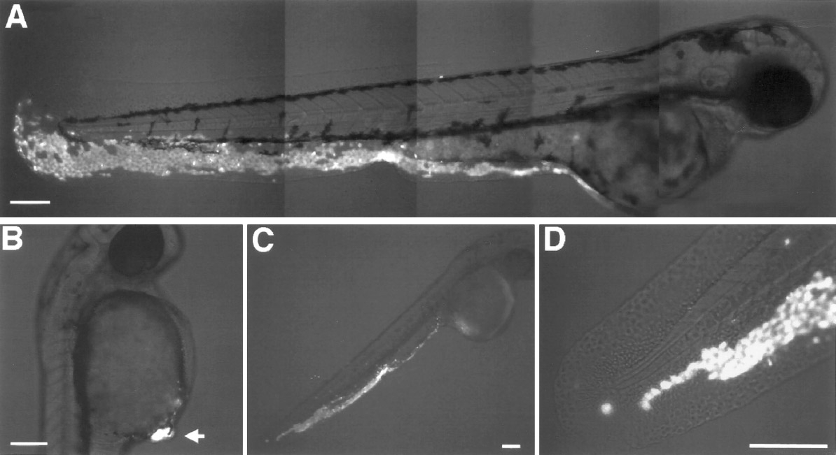
Fig. 3 The different behaviors of presumptive hindbrain progenitors transplanted at different times to identical positions in 6 h hosts. (A) Presumptive hindbrain progenitors (white) grafted at 6 h to the ventral epiblast region of a 6-h host integrate seamlessly to give rise to trunk and tail epidermis. (B) Presumptive hindbrain progenitors (white; arrow) grafted at 80% epiboly to a 6-h host form a small condensation of cells that does not integrate into the host. (C, closeup in D) Presumptive epidermal progenitors (white) grafted at 80% epiboly to a 6-h host integrate well into the host and gave rise to epidermis. Scale bar: all 100 μm.
Reprinted from Developmental Biology, 197, Woo, K. and Fraser, S.E., Specification of the hindbrain fate in the zebrafish, 283-296, Copyright (1998) with permission from Elsevier. Full text @ Dev. Biol.