
|
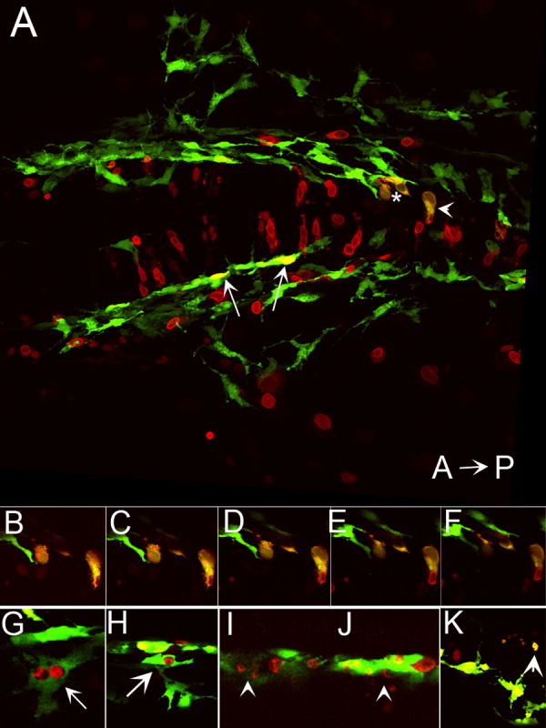
Fig. 8 Ectopic expression of foxd3 promotes cell death in kit mutants. A: Confocal stack image showing a dorsal view of the posterior head of a 28 hours postfertilization (hpf) kitw34/w34 embryo transgenic for mi:gfp and also expressing myc-foxd3 (red) under the control of a heat shock promoter. Expression of myc-foxd3 was induced at 19 hpf. Both wild-type (arrows) and fragmenting (asterisk and arrowhead) melanophores are observed. B-F: Confocal slices showing fragmenting cells indicated by asterisk and arrowhead in A. Note the presence of Foxd3-positive fragments within the GFP+ melanophores. G,H: Confocal slices showing additional examples of GFP/Foxd3+ cells designated as normal melanophores. I-K: Confocal slices showing examples of GFP/Foxd3+ cells designated as fragmenting melanophores (arrowheads).