
|
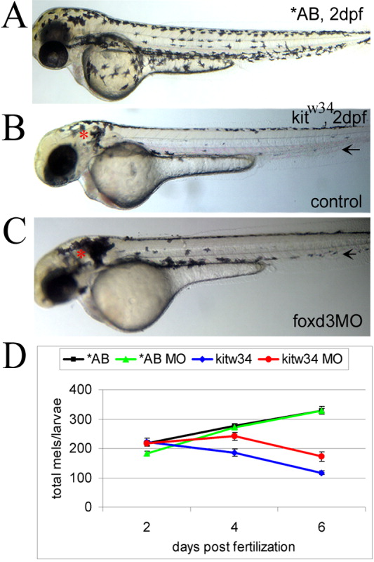
Fig. 2 foxd3 loss-of-function reduces loss of melanophores in kitw34 zebrafish. A: In 2 days postfertilization (dpf) wild-type zebrafish (AB), melanophores have migrated extensively, anteriorly to the head and ventrally over the yolk. B: In 2 dpf kitw34 zebrafish, melanophores are specified, but largely fail to migrate from sites of origin: the dorsal trunk and area caudal to the otic vesicle (red asterisk). C: kitw34 zebrafish injected with foxd3 morpholino oligonucleotide (MO) have more melanophores, especially apparent near the otic vesicle (foxd3MO, red asterisk). Both the ventral and lateral stripes show an increase in melanophores (black arrows). D: Line graph showing counts of total melanophores in 2, 4, and 6 dpf AB or kitw34 larvae, either uninjected (control) or injected with foxd3 MO. A significant change in the number of melanophores is observed in kitw34 mutants after injection of foxd3 MO (red) as compared to kitw34 uninjected controls (blue; P < 0.0001 by two-way analysis of variance (ANOVA); 7-11 fish per time point), whereas AB foxd3 morphants (green) show no significant difference as compared to uninjected AB controls (black; P = 0.49 by two-way ANOVA; 9-13 fish per time point).