
|
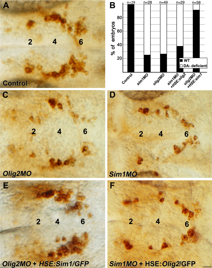
Fig. 5 Olig2 acts upstream to Sim1 in the control of DA specification. A, C-F: High-resolution micrographs (dorsal view, anterior to the left) displaying representative images of embryos from each treatment. At 52 hr post fertilization (hpf), embryos were fixed and subjected to immunostaining with an anti-TH antibody. Embryos were injected with sim1 MO (D,F) or olig2 MO (C,E). For the rescue analysis, embryos were injected with heat shock-inducible plasmid constructs containing either the sim1 (HSE:Sim1/GFP; E) or olig2 (HSE:Olig2/GFP; F) coding sequence. Expression of sim1 and olig2 was induced by shifting the embryos to the permissive temperature as described in the Experimental Procedures section. The amount of injected DNA HSE constructs in Olig2 and Sim1 gain-of-function experiments was determined by titrating the minimal doses of DNA that induce efficient ectopic RNA and GFP expression. B: Stacked bar chart representing the percentage of embryos displaying aberrant DA phenotype (denoted “DA- deficient”). Effects of the various genetic perturbations on DA neurons were scored and analyzed as described in the Experimental Procedures section. The number of embryos analyzed (n) is shown above. The major diencephalic DA groups 2, 4, and 6 are indicated. Scale bar = 25 μm.