
Image
|
Figure Caption
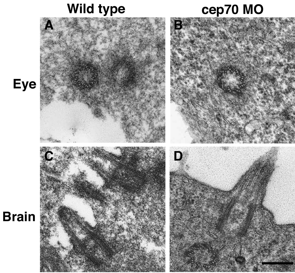
Fig. 8 Micrographs of basal bodies and cilia from WT and cep70 morphant zebrafish embryos. An eye centriole and brain cilium from 24 h.p.f. WT (A, C) and morphant (B, D) are shown. Scale bar: 250 nm
Figure Data
Acknowledgments
This image is the copyrighted work of the attributed author or publisher, and
ZFIN has permission only to display this image to its users.
Additional permissions should be obtained from the applicable author or publisher of the image.
Open Access.
Full text @ BMC Cell Biol.