
|
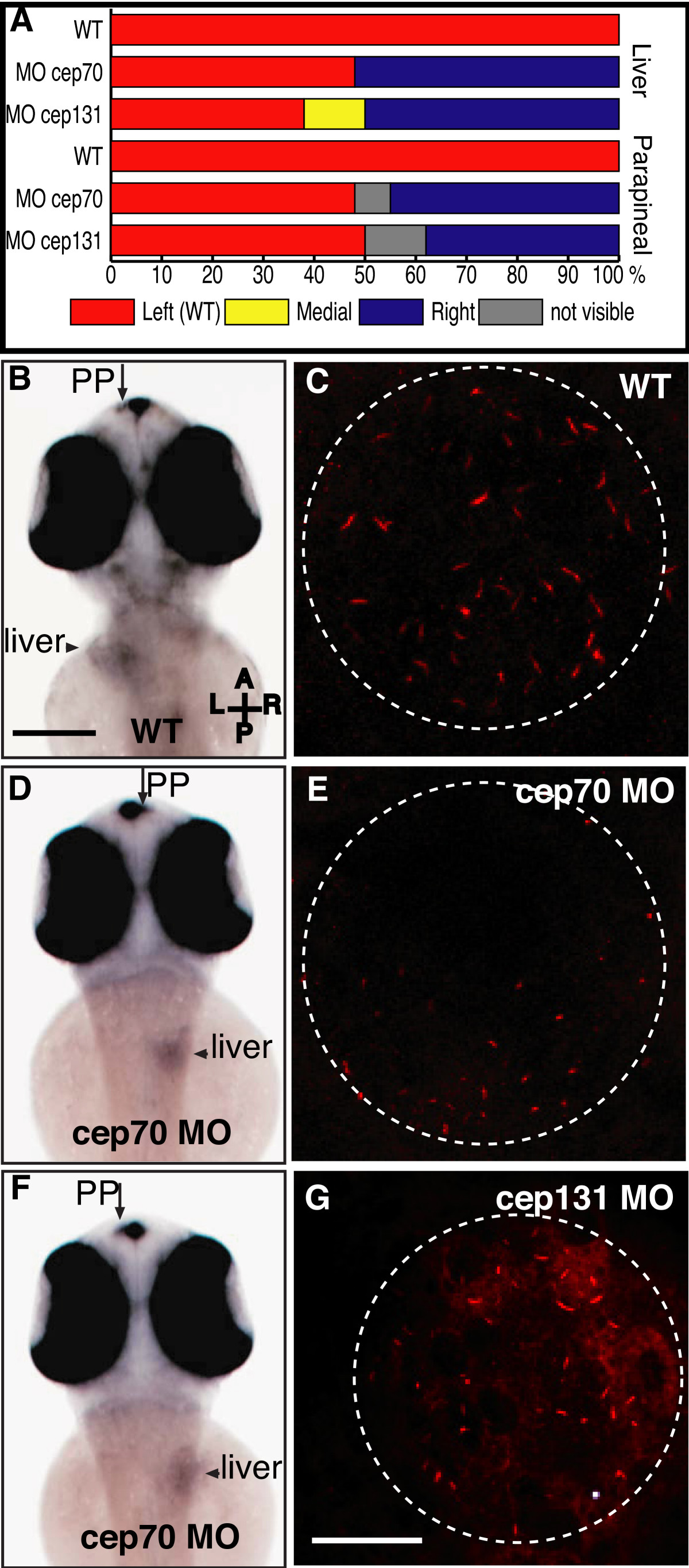
Fig. 5 Asymmetry in cep70 and cep131 morphants. (A) Randomisation of asymmetry of liver and parapineal in cep70 (n = 27) cep131 (n = 16) compared to WT (n = 21) where both are on the left hand side. (B, D, F) Position of asymmetrically placed organs in cep70 morphants compared to WT, at 60 h.p.f., assayed by wholemount in situ hybridisation for otx5 and fkd2 which label the parapineal (PP) and liver. In WT (B), both are placed on the left hand side. In cep70 morphants, this is randomised but not concordantly: the embryo in (D) has the position of both liver and parapineal inverted whereas the embryo in (F) has them on opposite sides – liver inverted, parapineal normal. Scale bar: 250 μm. Compass shows embryo orientation in which A = anterior, P = posterior, L = left and R = right. (C, E and G) Cilia in Kupffer's vesicle in WT, cep70 and cep131 morphants. Cilia are much shorter in the morphant embryos. Dashed circle outlines the vesicle. Scale bar: 20 μm.