
|
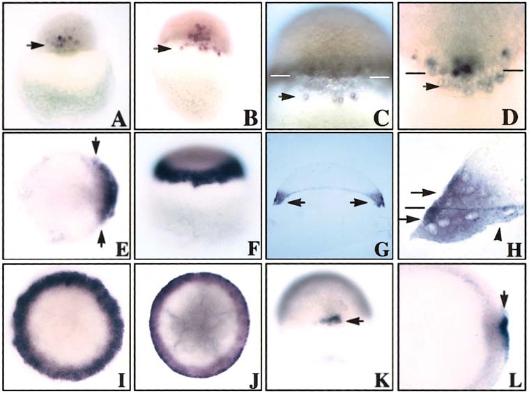
Fig. 2 Spatiotemporal expression pattern of znr2 by whole-mount in situ hybridization. (A) 512-cell/high stage. Arrow points to expression in dorsal blastomeres. Dorsal side view. (B–D) Oblong stage. Note localization of transcripts to the dorsal blastoderm and dorsal YSL. Arrows indicate expression around nuclei in YSL. Bars in C and D denote the blastoderm/yolk boundary. (E) Sphere stage. Note expansion, as shown by arrows, of the dorsal expression domain as znr2 transcripts begin to encircle the margin. Animal view. (F) Dome stage. Note expression around entire margin. Side view. (G) Dome stage. Section analysis shows znr2 expression in the margin. (H) Dome stage. Higher magnification of G. Top arrow shows expression in the deep and superficial cells of the blastoderm, the line marks the blastoderm/YSL boundary, the bottom arrow shows expression in the E-YSL, and the arrowhead points to expression in the I-YSL. (I) Fifty percent epiboly. Animal view. (J) Germ ring. Animal view. (K and L) Shield stage. Note expression in superficial dorsal forerunner cells of the dorsal shield. Dorsal and animal view, respectively.
Reprinted from Developmental Biology, 204, Erter, C.E., Solnica-Krezel, L., and Wright, C.V.E., Zebrafish nodal-related 2 encodes an early mesendodermal inducer signaling from the extraembryonic yolk syncytial layer, 361-372, Copyright (1998) with permission from Elsevier. Full text @ Dev. Biol.