
Image
|
Figure Caption
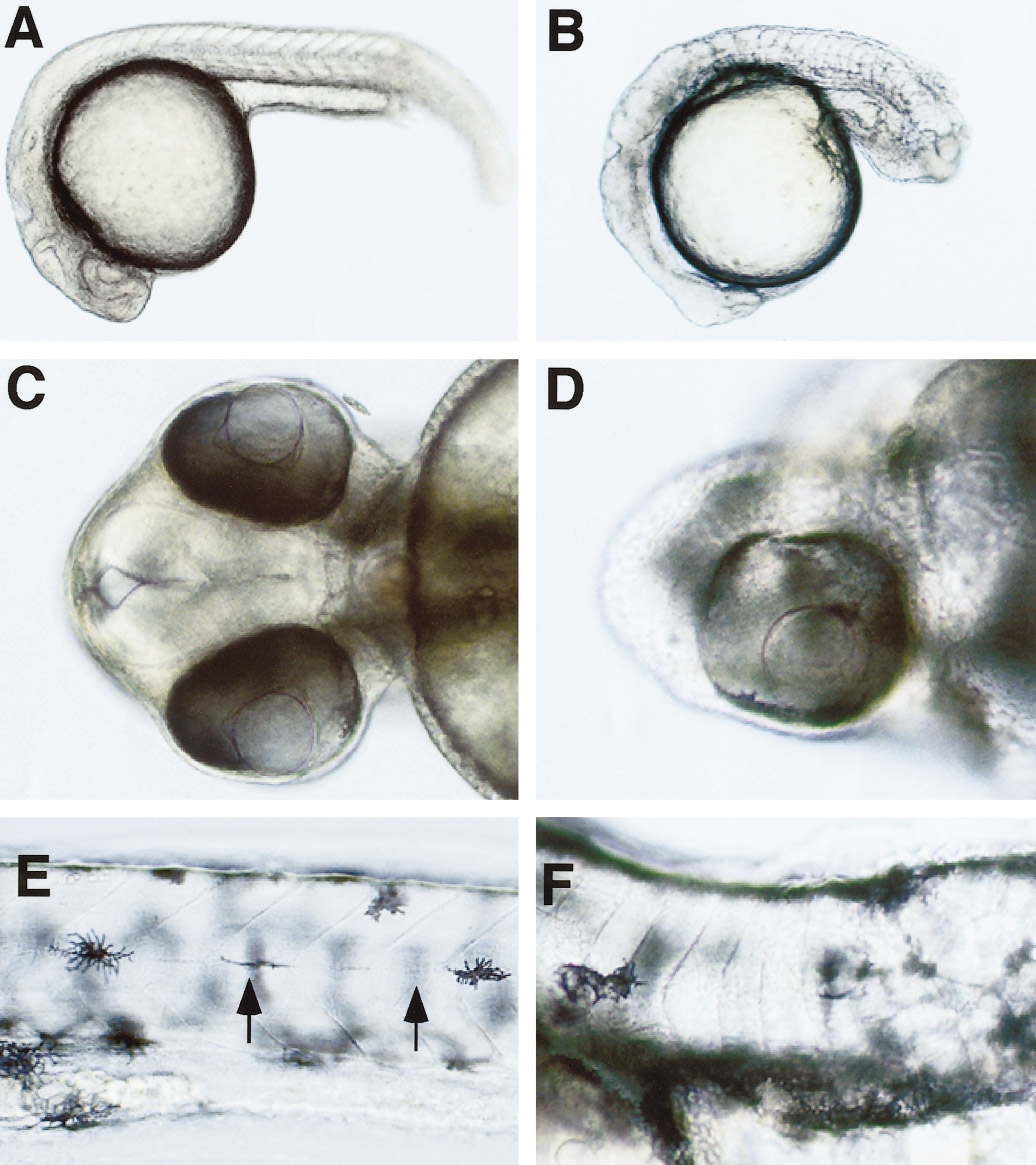
Fig. 1 Morphology of Ptx-injected embryos. (A, C, E) uninjected control, (B, D, F) Ptx-injected. (A, B) 30 hpf, lateral view; the injected embryo displays an altered shape of the head, blocky somites, and a generally disorganized tail. (C, D) 72 hpf, ventral view on head; the injected embryo displays cyclopia. (E, F) 96 hpf, lateral view on somites in trunk at level of anus; the somites of the injected embryo lack the chevron shape and the horizontal myoseptum (indicated in E by arrows). hpf, hours after fertilization.
Acknowledgments
This image is the copyrighted work of the attributed author or publisher, and
ZFIN has permission only to display this image to its users.
Additional permissions should be obtained from the applicable author or publisher of the image.
Reprinted from Developmental Biology, 194, Hammerschmidt, M. and McMahon, A.P., The effect of pertussis toxin on zebrafish development: a possible role for inhibitory G-proteins in hedgehog signaling, 166-171, Copyright (1998) with permission from Elsevier. Full text @ Dev. Biol.