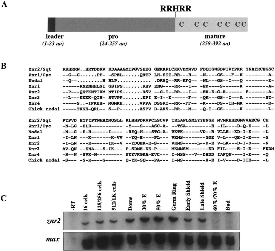
Fig. 1 Structure and expression of Znr2. (A) The deduced Znr2 sequence contains a hydrophobic signal sequence (leader), pro and mature ligand domains. Seven conserved cysteine residues in the ligand domain, and the RRHRR proteolytic cleavage site, are indicated. (B) Comparison of mature regions of the known Nodal-related ligands. Znr2/Sqt is 55, 48, 55, 50, 47, 45, and 55% identical to Znr1/Cyc, mouse Nodal, Xenopus Xnr1-4, and chick Nodal, respectively. Dashes represent identical residues, periods represent gaps. (C) RT-PCR analysis during development (stages above lanes) for znr2 expression. max was assayed as a loading control. Abbreviation: E, epiboly.
Reprinted from Developmental Biology, 204, Erter, C.E., Solnica-Krezel, L., and Wright, C.V.E., Zebrafish nodal-related 2 encodes an early mesendodermal inducer signaling from the extraembryonic yolk syncytial layer, 361-372, Copyright (1998) with permission from Elsevier. Full text @ Dev. Biol.

