
Fig. S4
- ID
- ZDB-IMAGE-090320-77
- Publication
- Jing et al., 2009 - Wnt signals organize synaptic prepattern and axon guidance through the zebrafish unplugged/MuSK receptor
- All Figures
- Figures for Jing et al., 2009
|
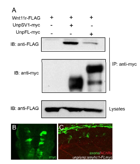
Fig. S4 Coimmunoprecipitation of UnpFL with Wnt11r in 293T cells and analysis of the Tg(smyhc1:UnpFLmyc) embryos. (A) 293T cells were cotransfected with Wnt11r-FLAG and UnpSV1-myc or UnpFL-myc. Whole cell lysates (WCL) were subjected to anti-FLAG immunoblotting (IB) to determine the expression of Wnt11r-FLAG (lower panel). The Wnt11r-FLAG binding was assessed by IB of the antimyc immunoprecipitate (upper panel). The amount of UnpFL or UnpSV1proteins were examined by anti-myc western blotting. Wnt11r-FLAG protein coimmunoprecipitated significantly better with UnpSV1 compared to UnpFL. (B) Cross-sectional view of a 17 hpf Tg embryo (smhyc1:unpFLmyc) stained with anti-myc. (C) Expression of UnpFL in adaxial cells failed to restore prepatterned AChRs (red) in unplugged embryos.