
Fig. S2
- ID
- ZDB-IMAGE-090320-75
- Publication
- Jing et al., 2009 - Wnt signals organize synaptic prepattern and axon guidance through the zebrafish unplugged/MuSK receptor
- All Figures
- Figures for Jing et al., 2009
|
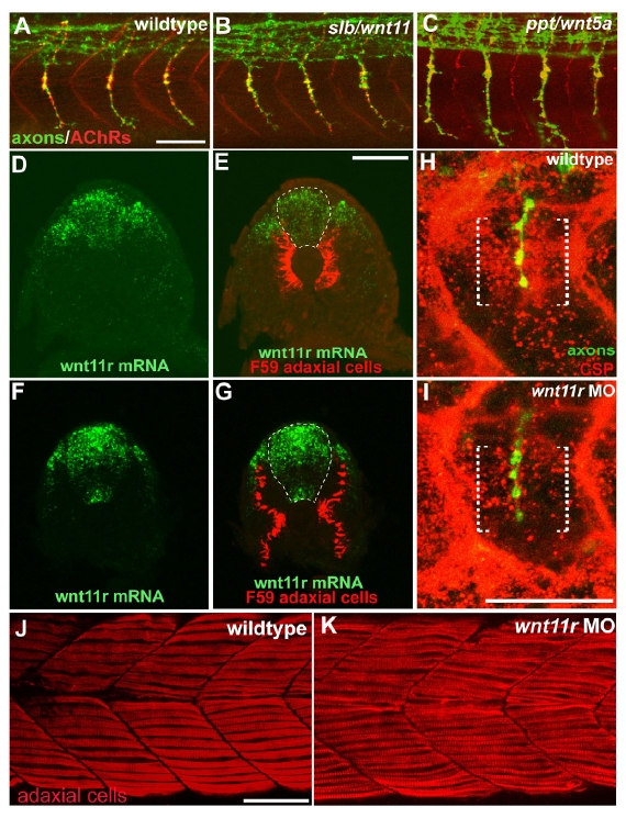
Fig. S2
wnt5/11 mutant analysis, wnt11r expression and stainings of wnt11r morphants.
(A) In 27 hpf wildtype embryos, neural AChR clusters (red, α-BTX) are apposed to primary motor axons (green, znp-1). In silberblick (slb/wnt11, B) and pipetail (ppt/wnt5a, C) mutants, axon pathfinding and neuromuscular synapses are unaffected. (D-G) Cross-section images from 20-somite stage wildtype embryos double stained for wnt11r mRNA (green) and adaxial cells (red, F59). In caudal segments (D and E), wnt11r is expressed in the spinal cord and in the dorso-lateral somites, just adjacent to pre-migratory adaxial cells. In the rostral segments (F and G), after the onset of adaxial cell migration, wnt11r expression is increased in the spinal cord. Dashed lines indicate the spinal cord. (H and I) Confocal images of embryos at 26-somite stage stained for CSPs (red) and motor axons (green). CSPs accumulate around the choice point (brackets) in wildtype embryos (H), but are reduced in wnt11r morphants (I). (J-K) Lateral views of adaxial fibers in wildtype and wnt11r MO-injected embryos. Scale bars: 50 μm.