
|
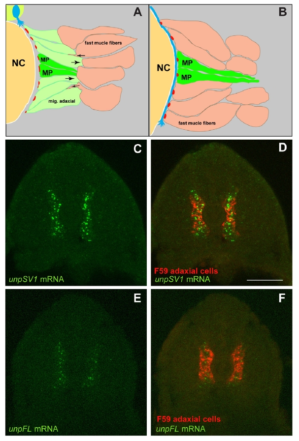
Fig. S1
Unplugged full-length (FL) and Splice Variant 1 (SV1) are differentially expressed.
(A-B) Schematic representation of AChR clustering. (A) Prior to the arrival of growth cones, migratory adaxial cells (light green) flank non-migratory adaxial or muscle pioneer cells (dark green, MP) and form a monolayer along the medial somite. Prepatterned AChR clusters (red) accumulate on the medial surface of all adaxial cells. As the first growth cones (blue) approaches, migratory adaxial fibers (light green) initiate their radial migration to the lateral surface of the somite, while fast muscle fibers (peachy) invade the space on the medial somite surface. NC, notochord. (B) Growth cones contact fast fibers and form neural en passant synapses. At the horizontal midline, growth cones contact muscle pioneers and incorporates prepatterned clusters into NMJs. (C-F) Confocal images of cross-sections from 20-somite stage wildtype embryos double stained for unplugged mRNA and F59 antibody, specific for adaxial cells. Just before the first motor axon exit from the spinal cord, unplugged SV1 is highly expressed in all premigratory adaxial cells (C-D), while unplugged FL is expressed at a much lower level (EF) compared to unplugged SV1. Scale bar: 50 μm.