
Image
|
Figure Caption
Fig. S6
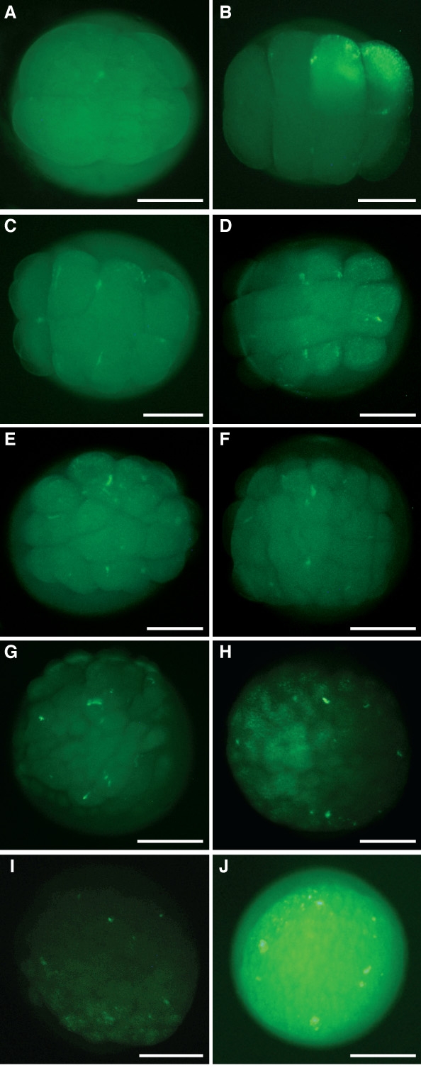
Buc-GFP Overexpression Generates Additional Fluorescent Spots
(A-J) Living embryos, animal view after injection of 200 pg buc-GFP mRNA at the one cell stage shown at the 4-cell (A) 8-cell (B) 8- to 16-cell (C) 16-cell (D) 16- to 32- cell (E) 32-cell (F) 64-cell (G) 128-cell (H) high (I) and oblong stage (J). Note that the oblong stage embryo (J) is overexposed, since the fluorescence is slowly decreasing and becomes invisible around dome stage. Scale bars: 200 μm
Acknowledgments
This image is the copyrighted work of the attributed author or publisher, and
ZFIN has permission only to display this image to its users.
Additional permissions should be obtained from the applicable author or publisher of the image.
Full text @ Curr. Biol.