
|
Fig. S2
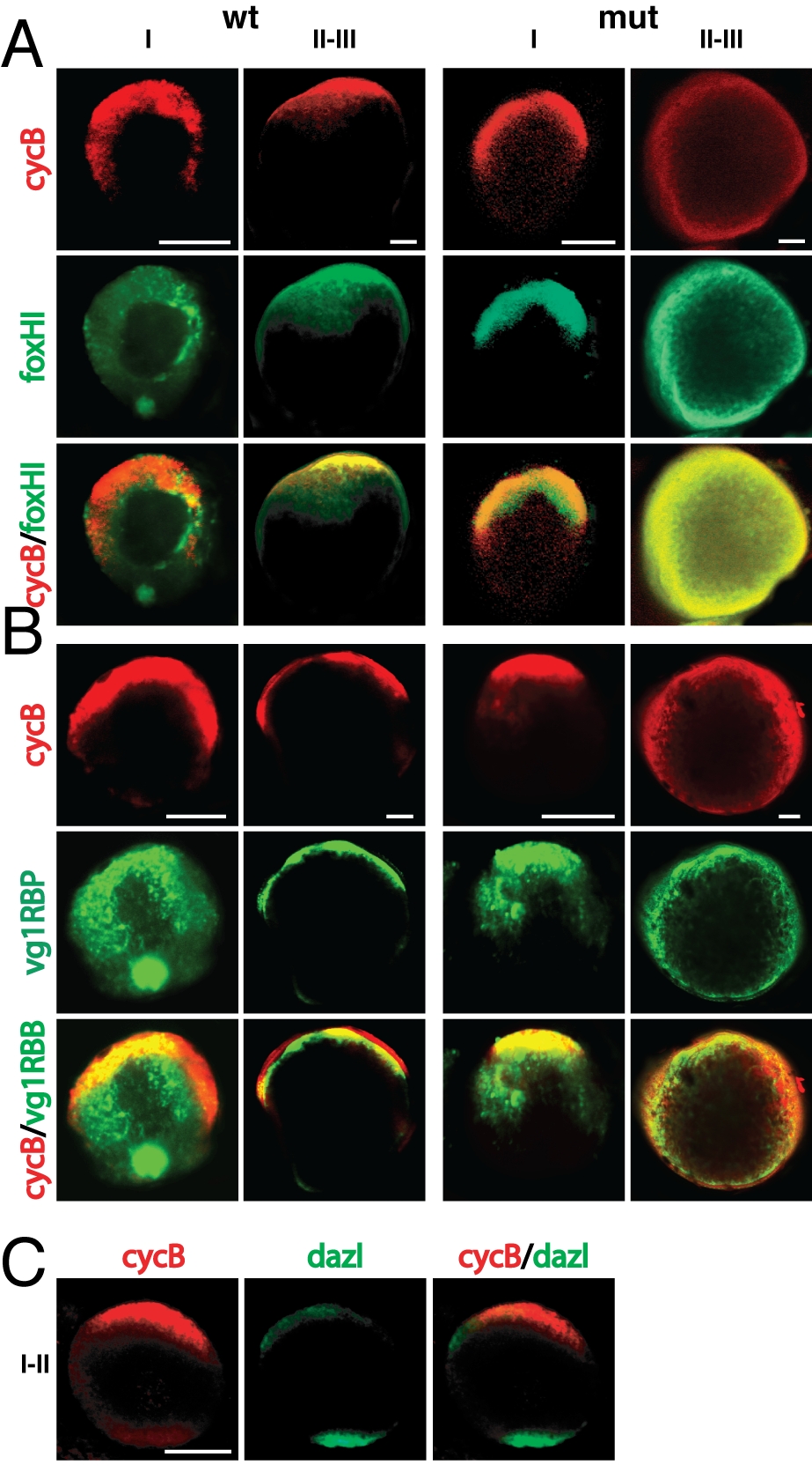
cyclinB, foxH1, and vg1RBP mRNA Localization in Wild-Type and buc Mutants
(A, B) Whole-mount in situ hybridization with cyclinB (red) and foxH1 mRNA (green) (A) or vg1RBP (green) (B) in stage I and II-III oocytes. Animal to the top. (A) CycB mRNA stays localized at the animal pole in wild type as well as early mutant oocytes. Some of these panels are double stainings from the single channels displayed in Fig. 1 to confirm the localization of the used molecular markers. In wild type foxH1 (A) and vg1RBP mRNA (B) are localized to the Balbiani body at stage I and animal at stage II-III. In buc oocytes foxH1 (A) and vg1RBP (B) are localized animal at stage I. Scale bar: 25 μm for stage I, 50 μm for stage II-III.
(C) CycB (red) and dazl mRNA (green) are localized at opposite poles at the end of stage I. Scale bar: 50 μm.