
|
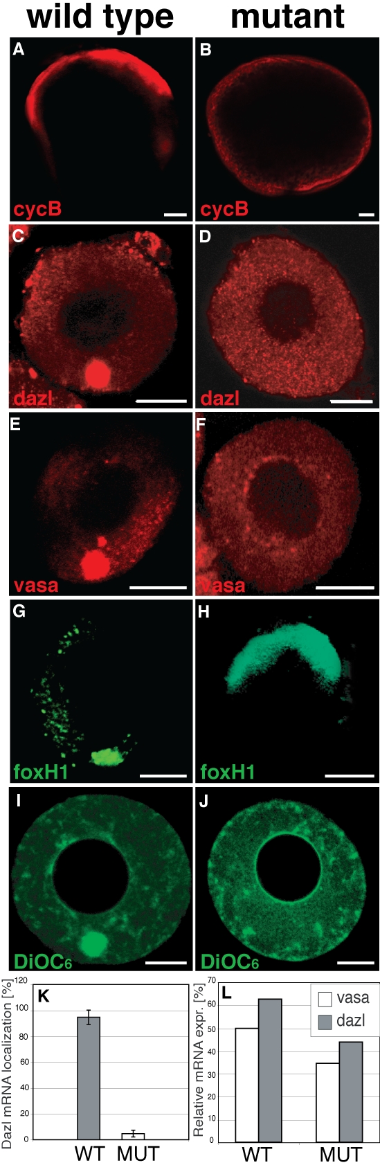
Fig. S1
Egg Polarity and Germ Plasm Formation in Early Bucky Ball Oocytes
(A-H) Fluorescent whole-mount in situ hybridization of stage III (A, B) and early Ib oocytes (C-H). cyclinB (A, B), dazl (C, D), vasa (E, F) (red) and foxH1 (G, H) (green) in wild-type (A, C, E, G) and buc p106re mutant stage I oocytes (B, D, F, H).
(I, J) Living staining of mitochondria and endoplasmic reticulum with DiOC6 (green) in wt (I) and buc p106re mutant oocytes (J). Note the absence of signal at the vegetal pole in mutants. Animal pole to the top. Scale bar: 50 μm (A, B), 25μm (C-J).
(K) Quantification of oocytes with localized dazl mRNA. Note dazl mRNA localization in wild type (WT) (94.7±5.5%; n=385; grey bar) but not in buc p106re mutants (MUT) (4.8±2.6%; n=402; white bar). Error bars indicate the standard deviation of the average (at least three independent experiments).
(L) Quantitative real-time PCR measuring the relative concentration of vasa (white) and dazl (grey) mRNA in whole ovaries from wt and buc p106re mut females.